Principles of cell signaling - UT Southwestern
Principles of cell signaling - UT Southwestern
Principles of cell signaling - UT Southwestern

You also want an ePaper? Increase the reach of your titles
YUMPU automatically turns print PDFs into web optimized ePapers that Google loves.
39057_ch14_<strong>cell</strong>bio.qxd 8/28/06 5:11 PM Page 617<br />
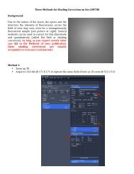
Structure <strong>of</strong> rhodopsin<br />
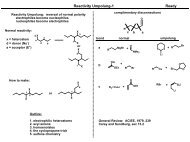
Heterotrimeric G protein structure<br />
CYTOPLASM<br />
MEMBRANE<br />
Retinal<br />
FIGURE 14.23 The figure shows the crystal structure<br />
<strong>of</strong> the GPCR rhodopsin. Each membrane-spanning helix<br />
is a different color; most structures on the cytoplasmic<br />
face are not shown. The retinal chromophore is<br />
shown within the helix bundle. GPCR sequence similarity<br />
separates the mammalian GPCRs into at least four<br />
structural families that are so diverse that there may<br />
be little sequence similarity among the classes. Within<br />
a family, similarity is greatest in the membrane-spanning<br />
helices, less in the interhelical loops, and least in<br />
the N- and C-terminal domains and in the cytoplasmic<br />
loop that connects spans five and six. Regardless, the<br />
generalizations about functional domains in receptors<br />
seem to hold true within different families. GPCRs frequently<br />
form dimers, occasionally heterodimers, and<br />
dimerization can be crucial for function. Structure generated<br />
from Protein Data Bank file 1F88.<br />
The heterotrimeric G proteins to which<br />
GPCRs are coupled are composed <strong>of</strong> a nucleotide-binding<br />
Gα subunit and a Gβγ subunit<br />
dimer, as illustrated in FIGURE 14.24. The structure<br />
<strong>of</strong> the trimer and each subunit is known for<br />
several states <strong>of</strong> activation and in complex with<br />
several interacting proteins. A Gαβγ heterotrimer<br />
is named according to its α subunit, which largely<br />
defines the G protein’s selectivity among receptors.<br />
Each subunit also regulates a distinct group<br />
<strong>of</strong> effector proteins.<br />
Gα subunits are globular, two-domain proteins<br />
<strong>of</strong> 38-44 kDa. The GTP-binding domain<br />
belongs to the GTP-binding protein superfamily<br />
that includes the small, monomeric G proteins<br />
(such as Ras, Rho, Arf, Rab; see 14.23 Small,<br />
monomeric GTP-binding proteins are multiuse<br />
switches) as well as the GTP-binding translational<br />
initiation and elongation factors. A second domain<br />
modulates GTP binding and hydrolysis.<br />
Gα subunits are only slightly hydrophobic, but<br />
they are predominantly membrane-associated<br />
FIGURE 14.24 The structure <strong>of</strong> the nonactivated G i<br />
heterotrimer,<br />
the G protein that is responsible for inhibition <strong>of</strong><br />
adenylyl cyclase and for most G-mediated <strong>signaling</strong>, is<br />
shown with each subunit colored as shown. GDP is shown<br />
bound to the G i<br />
subunit. Structure generated from Protein<br />
Data Bank file 1GP2.<br />
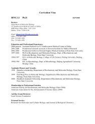
G<br />
protein<br />
Gs<br />
Golf<br />
Gi (3)<br />
Go<br />
Gz<br />
Ggus<br />
Gt (2)<br />
Gq (4)<br />
G 12<br />
G 13<br />
Adenylyl cyclase<br />
EFFECTOR PROTEIN<br />
K+channel, PI 3-kinase<br />
Other cation channel<br />
Rho GEF<br />
G protein targets<br />
Stimulated<br />
Cyclic GMP phosphodiesterase<br />
Phospholipase-Cβ<br />
Inhibited<br />
Adenylyl cyclase<br />
FIGURE 14.25 G protein-regulated effectors do not share structural<br />
similarities. They may be ion channels or membrane spanning<br />
enzymes in the plasma membrane, peripheral proteins on the<br />
inner face <strong>of</strong> the membrane, or fundamentally soluble proteins<br />
that can bind to G subunits. The chart shows the major groups<br />
<strong>of</strong> G proteins, sorted according to sequence similarity, and some<br />
<strong>of</strong> the effectors that they are known to regulate.<br />
because <strong>of</strong> constitutive N-terminal fatty acylation<br />
and because they bind to the membraneattached<br />
Gβγ subunits. Mammals have 16 Gα<br />
genes that are grouped in subfamilies according<br />
to similar sequence and function (e.g., s, i, q,<br />
and 12). These subfamilies are listed in FIGURE<br />
14.25.<br />
Gβ and Gγ subunits associate irreversibly<br />
soon after translation to form stable Gβγ dimers,<br />
which then associate reversibly with a Gα. Gβ<br />
subunits are 35 kDa proteins composed <strong>of</strong> seven<br />
14.20 G protein <strong>signaling</strong> modules are widely used and highly adaptable 617