Supplementum 3+4/2007 - SpoleÄnost pro pojivové tkánÄ›
Supplementum 3+4/2007 - SpoleÄnost pro pojivové tkánÄ›
Supplementum 3+4/2007 - SpoleÄnost pro pojivové tkánÄ›

You also want an ePaper? Increase the reach of your titles
YUMPU automatically turns print PDFs into web optimized ePapers that Google loves.
ar spine (6). MRI showed evident collapse<br />
of the vertebrae because of tuberculous<br />
destruction and paravertebral abscess.<br />
Neurological deficits were found in 6 patients.<br />
One case was graded B, two cases<br />
were graded C, and three cases were graded<br />
D according to Frankel classification.<br />
Technique<br />
Before surgery, patients received standard<br />
anti-tuberculosis chemotherapy for 2<br />
to 3 weeks. Retroperitoneal or extrapleural<br />
ap<strong>pro</strong>ach was chosen according to the<br />
tuberculosis lesion segment. Anterior radical<br />
debridement, iliac or rib autografting<br />
and anterior plating was used<br />
Postoperative management<br />
Anti-tuberculosis chemotherapy was<br />
continued for at least 9 months, and the<br />
patients were supported with thoracolumbosacral<br />
orthosis for 6 months after surgery.<br />
All patients were followed up for an<br />
average of 18 months. On each assessment,<br />
data related to drug regimen and its side<br />
effects if any, abscess or sinus formation,<br />
im<strong>pro</strong>vement of back pain and tenderness<br />
were recorded.<br />
Postoperative neurological assessment<br />
was reported and compared with the preoperative<br />
state The activity of the disease<br />
was assisted by ESR at monthly intervals<br />
for the first 3 months, then once every<br />
3 months during the first year, and every<br />
6 months until the final follow up.<br />
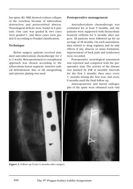
Anteroposterior and lateral radiographs<br />
of the spine were obtained each visit<br />
Figure 3. Follow up X-rays 6 months after surgery<br />
300 The 9 th Prague-Sydney-Lublin Symposium