LATVIA UNIVERSITY OF AGRICULTURE - Latvijas ...
LATVIA UNIVERSITY OF AGRICULTURE - Latvijas ...
LATVIA UNIVERSITY OF AGRICULTURE - Latvijas ...
- No tags were found...
You also want an ePaper? Increase the reach of your titles
YUMPU automatically turns print PDFs into web optimized ePapers that Google loves.
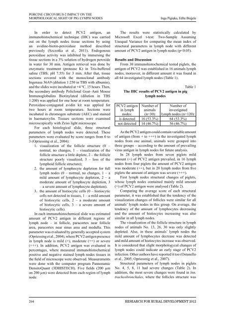
PORCINE CIRCOVIRUS-2 IMPACT ON THEMORPHOLOGICAL SIGHT <strong>OF</strong> PIG LYMPH NODESInga Pigiņka, Edīte BirģeleIn order to detect PCV2 antigen, animmunohistochemical technique (IHC) was carriedout on the lymph nodes tissue sections by usingan avidine-biotin-peroxidase method describedpreviously (Szczotka et al., 2011). Endogenousperoxidase activity was inhibited by immersing thetissue sections in a 3% solution of hydrogen peroxidein water for 30 min. Antigen retrieval was done byenzymatic treatment (protease K) in Tris-bufferedsaline (TBS; pH 7.55) for 3 min. After that, tissuesections covered with the monoclonal antibodyIngenasa 36A9 (dilution 1:250 in TBS with albumin),and the slides were incubated at +4 °C, 15 hours. Then,the secondary antibody Policlonal Goat–Anti MouseImmunoglobulins Biotinylated (dilution in TBS1:200) was applied for one hour at room temperature.Peroxidase-conjugated avidin kit was applied fortwo hours at room temperature. Sections wereincubated in chromogen substrate (AEC) and stainedin haematoxylin. Tissues sections were examinedmicroscopically with Zeiss light microscope.For each histological slide, three structuralparameters of lymph nodes were detected. Theseparameters were evaluated by score ranges from 0 to3 (Opriessing et al., 2004):1. visualization of the follicle structure (0 –normal, no changes, 1 – visualization of thefollicle structure a little deplete, 2 – the folliclestructure poorly visualized, 3 – loss of thelymphoid follicle structure);2. the amount of lymphocyte depletion for fulllymph nodes (0 – normal, no changes, 1 – amild amount of lymphocyte depletion, 2 – amoderate amount of lymphocyte depletion, 3– a severe amount of lymphocyte depletion);3. the amount of histiocytic cells (0 – histiocyticcells not detected in tissues, 1 – a mild amountof histiocytic cells, 2 – a moderate amountof histiocytic cells, 3 – a severe amount ofhistiocytic cells).In each immunohistochemical slide was estimatedamount of PCV2 antigen in different regions oflymph node – in follicle, paracortex near folliclearea, paracortex near sinus area and medulla. Thisparameter was evaluated by generally accepted system(Opriessing et al., 2004), where PCV2 antigen presencein lymph node is mild (+), moderate (++) or severe(+++). In addition, PCV2 antigen was evaluated inpercentages, where measured immunohistochemicalpositive and negative stained lymph nodes tissues inthe field of microscope were observed. Measurementswere done with the computer program PannoramicDensitoQuant (3DHISTECH). Five fields (200 µmon 200 µm) were detected from each region of lymphnode.The results were statistically calculated byMicrosoft Excel t-test: Two-Sample AssumingUnequal Variance for comparing the mean index ofstructural parameters in lymph node with differentamount of PCV2 antigen in lymph nodes (p


