LATVIA UNIVERSITY OF AGRICULTURE - Latvijas ...
LATVIA UNIVERSITY OF AGRICULTURE - Latvijas ...
LATVIA UNIVERSITY OF AGRICULTURE - Latvijas ...
- No tags were found...

Create successful ePaper yourself
Turn your PDF publications into a flip-book with our unique Google optimized e-Paper software.
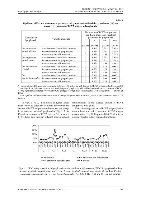
Inga Pigiņka, Edīte BirģelePORCINE CIRCOVIRUS-2 IMPACT ON THEMORPHOLOGICAL SIGHT <strong>OF</strong> PIG LYMPH NODESTable 5Significant difference in structural parameters of lymph node with mild (+), moderate (++) andsevere (+++) amount of PCV2 antigen in lymph nodeThe name oflymph nodeValued parametersThe amount of PCV2 antigen andsignificant changes in structuralparameters in lymph node- + ++ +++lnn. inguinalessuperf. sinisterlnn. inguinalessuperf. dexterlnn. mesentericicranialelnn.tracheobronchales(n=56) (n=28) (n=16) (n=20)visualization of the follicle structure 0 1.86* 2.75^ 2.80¬decrease amount of lymphocytes 0 1.43* 2.25^ 2.20¬increase amount of histiocytes 0 1.43* 1.50 2.00¬visualization of the follicle structure 0 1.86* 2.75^ 2.80¬decrease amount of lymphocytes 0 1.43* 2.25^ 2.20¬increase amount of histiocytes 0 1.43* 1.50 2.50°¬visualization of the follicle structure 0 2.00* 2.25 3.00°¬decrease amount of lymphocytes 0 1.57* 1.75 2.80°¬increase amount of histiocytes 0 1.57* 1.50 2.40°¬visualization of the follicle structure 0 2.14* 2.00 3.00°¬decrease amount of lymphocytes 0 1.57* 2.00 3.00°¬increase amount of histiocytes 0 1.57* 1.75 2.60°¬* – the significant difference between structural changes of lymph node with 0 amount of PCV2 antigen and mild amount (+)^ – the significant difference between structural changes of lymph node with mild (+) and moderate (++) amount of PCV2° – the significant difference between structural changes of lymph node with moderate (++) and severe (+++) amount ofPCV2¬ – the significant difference between structural changes of lymph nodes with mild (+) and severe (+++) amount of PCV2in themTo view a PCV2 distribution in lymph nodesfrom follicle to other part of lymph node better, theamount of PCV2 antigen was reflected as a percentagein separate structures of lymph nodes (Fig. 1, 2, 3).Considering amount of PCV2 antigen (%) measuredby five fields from each part of lymph nodes, graphicalrepresentations in the average amount of PCV2antigen (%) were given.First, the average amount of PCV2 antigen (%) forseven animals with mild (+) amount of PCV2 antigenwas evaluated (Fig. 1). It appeared that PCV2 antigenis mainly located in the lymph nodes follicles.20%15%10%5%0%Lnn.ALnn.BLnn.CLnn.DLnn.ALnn.BLnn.CLnn.DLnn.ALnn.BLnn.CLnn.DLnn.ALnn.BLnn.CLnn.DLnn.ALnn.BLnn.CLnn.DLnn.ALnn.BLnn.CLnn.DLnn.ALnn.BLnn.CLnn.DNo.4 No.5 No.8 No.11 No.13 No.26 No.30folliculeparacortex near sinus areaparacortex near follicule areamedullaFigure 1. PCV2 antigen location in lymph nodes mainly with mild (+) amount of PCV2 in lymph nodes: Lnn.A – lnn. inguinales superficiales sinister, Lnn. B – lnn. inguinales superficiales sinister dexter, Lnn. C – lnn.mesenterici cranial and Lnn. D – lnn. tracheobronchales. No. 4, 5, 8, 11, 13, 26 and 30 – animal number.Research for Rural Development 2012207