View - DSpace UniPR
View - DSpace UniPR
View - DSpace UniPR

You also want an ePaper? Increase the reach of your titles
YUMPU automatically turns print PDFs into web optimized ePapers that Google loves.
Chapter 6<br />
6.2.2 Uptake experiments<br />
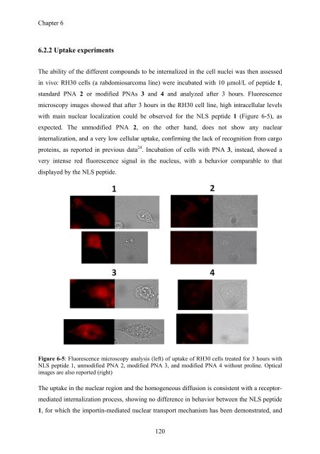
The ability of the different compounds to be internalized in the cell nuclei was then assessed<br />
in vivo: RH30 cells (a rabdomiosarcoma line) were incubated with 10 µmol/L of peptide 1,<br />
standard PNA 2 or modified PNAs 3 and 4 and analyzed after 3 hours. Fluorescence<br />
microscopy images showed that after 3 hours in the RH30 cell line, high intracellular levels<br />
with main nuclear localization could be observed for the NLS peptide 1 (Figure 6-5), as<br />
expected. The unmodified PNA 2, on the other hand, does not show any nuclear<br />
internalization, and a very low cellular uptake, confirming the lack of recognition from cargo<br />
proteins, as reported in previous data 24 . Incubation of cells with PNA 3, instead, showed a<br />
very intense red fluorescence signal in the nucleus, with a behavior comparable to that<br />
displayed by the NLS peptide.<br />
Figure 6-5: Fluorescence microscopy analysis (left) of uptake of RH30 cells treated for 3 hours with<br />
NLS peptide 1, unmodified PNA 2, modified PNA 3, and modified PNA 4 without proline. Optical<br />
images are also reported (right)<br />
The uptake in the nuclear region and the homogeneous diffusion is consistent with a receptormediated<br />
internalization process, showing no difference in behavior between the NLS peptide<br />
1, for which the importin-mediated nuclear transport mechanism has been demonstrated, and<br />
120