Coral Health and Disease in the Pacific: Vision for Action
Coral Health and Disease in the Pacific: Vision for Action
Coral Health and Disease in the Pacific: Vision for Action
Create successful ePaper yourself
Turn your PDF publications into a flip-book with our unique Google optimized e-Paper software.
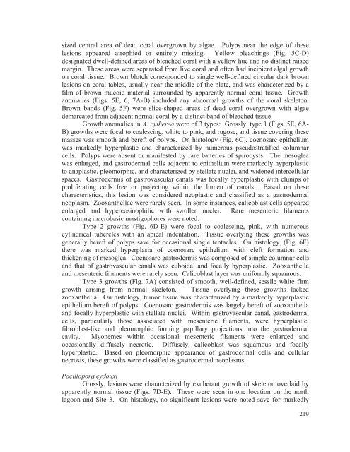
sized central area of dead coral overgrown by algae. Polyps near <strong>the</strong> edge of <strong>the</strong>selesions appeared atrophied or entirely miss<strong>in</strong>g. Yellow bleach<strong>in</strong>gs (Fig. 5C-D)designated dwell-def<strong>in</strong>ed areas of bleached coral with a yellow hue <strong>and</strong> no dist<strong>in</strong>ct raisedmarg<strong>in</strong>. These areas were separated from live coral <strong>and</strong> often had <strong>in</strong>cipient algal growthon coral tissue. Brown blotch corresponded to s<strong>in</strong>gle well-def<strong>in</strong>ed circular dark brownlesions on coral tables, usually near <strong>the</strong> middle of <strong>the</strong> plate, <strong>and</strong> was characterized by afilm of brown mucoid material surrounded by apparently normal coral tissue. Growthanomalies (Figs. 5E, 6, 7A-B) <strong>in</strong>cluded any abnormal growths of <strong>the</strong> coral skeleton.Brown b<strong>and</strong>s (Fig. 5F) were slice-shaped areas of dead coral overgrown with algaedemarcated from adjacent normal coral by a dist<strong>in</strong>ct b<strong>and</strong> of bleached tissueGrowth anomalies <strong>in</strong> A. cy<strong>the</strong>rea were of 3 types: Grossly, type 1 (Figs. 5E, 6A-B) growths were focal to coalesc<strong>in</strong>g, white to p<strong>in</strong>k, <strong>and</strong> rugose, <strong>and</strong> tissue cover<strong>in</strong>g <strong>the</strong>semasses was smooth <strong>and</strong> bereft of polyps. On histology (Fig. 6C), coenosarc epi<strong>the</strong>liumwas markedly hyperplastic <strong>and</strong> characterized by numerous pseudostratified columnarcells. Polyps were absent or manifested by rare batteries of spirocysts. The mesogleawas enlarged, <strong>and</strong> gastrodermal cells adjacent to epi<strong>the</strong>lium were markedly hyperplasticto anaplastic, pleomorphic, <strong>and</strong> characterized by stellate nuclei, <strong>and</strong> widened <strong>in</strong>tercellularspaces. Gastrodermis of gastrovascular canals was focally hyperplastic with clumps ofproliferat<strong>in</strong>g cells free or project<strong>in</strong>g with<strong>in</strong> <strong>the</strong> lumen of canals. Based on <strong>the</strong>secharacteristics, this lesion was considered neoplastic <strong>and</strong> classified as a gastrodermalneoplasm. Zooxan<strong>the</strong>llae were rarely seen. In some <strong>in</strong>stances, calicoblast cells appearedenlarged <strong>and</strong> hypereos<strong>in</strong>ophilic with swollen nuclei. Rare mesenteric filamentsconta<strong>in</strong><strong>in</strong>g macrobasic mastigophores were noted.Type 2 growths (Fig. 6D-E) were focal to coalesc<strong>in</strong>g, p<strong>in</strong>k, with numerouscyl<strong>in</strong>drical tubercles with an apical <strong>in</strong>dentation. Tissue overly<strong>in</strong>g <strong>the</strong>se growths wasgenerally bereft of polyps save <strong>for</strong> occasional s<strong>in</strong>gle tentacles. On histology, (Fig. 6F)<strong>the</strong>re was marked hyperplasia of coenosarc epi<strong>the</strong>lium with cleft <strong>for</strong>mation <strong>and</strong>thicken<strong>in</strong>g of mesoglea. Coenosarc gastrodermis was composed of simple columnar cells<strong>and</strong> that of gastrovascular canals was cuboidal <strong>and</strong> focally hyperplastic. Zooxan<strong>the</strong>lla<strong>and</strong> mesenteric filaments were rarely seen. Calicoblast layer was uni<strong>for</strong>mly squamous.Type 3 growths (Fig. 7A) consisted of smooth, well-def<strong>in</strong>ed, sessile white firmgrowth aris<strong>in</strong>g from normal skeleton. Tissue overly<strong>in</strong>g <strong>the</strong>se growths lackedzooxan<strong>the</strong>lla. On histology, tumor tissue was characterized by a markedly hyperplasticepi<strong>the</strong>lium bereft of polyps. Coenosarc gastrodermis was largely bereft of zooxan<strong>the</strong>lla<strong>and</strong> focally hyperplastic with stellate nuclei. With<strong>in</strong> gastrovascular canal, gastrodermalcells, particularly those associated with mesenteric filaments, were hyperplastic,fibroblast-like <strong>and</strong> pleomorphic <strong>for</strong>m<strong>in</strong>g papillary projections <strong>in</strong>to <strong>the</strong> gastrodermalcavity. Myonemes with<strong>in</strong> occasional mesenteric filaments were enlarged <strong>and</strong>occasionally diffusely necrotic. Diffusely, calicoblast was squamous <strong>and</strong> focallyhyperplastic. Based on pleomorphic appearance of gastrodermal cells <strong>and</strong> cellularnecrosis, <strong>the</strong>se growths were classified as gastrodermal neoplasms.Pocillopora eydouxiGrossly, lesions were characterized by exuberant growth of skeleton overlaid byapparently normal tissue (Figs. 7D-E). These were seen <strong>in</strong> one location on <strong>the</strong> northlagoon <strong>and</strong> Site 3. On histology, no significant lesions were noted save <strong>for</strong> markedly219


