

world cancer report - iarc
world cancer report - iarc
world cancer report - iarc

You also want an ePaper? Increase the reach of your titles
YUMPU automatically turns print PDFs into web optimized ePapers that Google loves.
SCREENING FOR ORAL CANCER<br />
SUMMARY<br />
> Oral <strong>cancer</strong> and its pre<strong>cancer</strong>ous<br />
lesions, including leukoplakia, can be<br />
readily detected by visual inspection of<br />
the oral cavity by trained health workers<br />
and doctors.<br />
> Population screening for oral <strong>cancer</strong><br />
results in the diagnosis of large numbers<br />
of oral pre<strong>cancer</strong>s and an<br />
increased proportion of early stage<br />
tumours. However, a reduction in incidence<br />
of and mortality from oral <strong>cancer</strong><br />
resulting from such interventions<br />
remains to be demonstrated.<br />
Oral lesions such as leukoplakia, erythroplakia<br />
and oral submucous fibrosis are<br />
pre<strong>cancer</strong>ous. A high risk of malignant<br />
transformation of such lesions has been<br />
established in follow-up studies. The proportion<br />
of oral <strong>cancer</strong>s that arise from<br />
pre-existing pre<strong>cancer</strong>ous lesions is variously<br />
<strong>report</strong>ed in the range of 30-80%.<br />
The natural history of these lesions is not<br />
as extensively documented as that of the<br />
precursors to cervical <strong>cancer</strong>. Thus, for<br />
example, it is not known whether the different<br />
types of leukoplakia and erythroplakia<br />
constitute a continuum similar to<br />
the different stages evident during the<br />
development of cervical intraepithelial<br />
neoplasia.<br />
Oral leukoplakia refers to uniform, flat,<br />
predominantly white lesions in the lining<br />
of the mouth that cannot be characterized<br />
as any other disease. White lesions<br />
with a smooth, corrugated or wrinkled<br />
surface are referred to as homogeneous<br />
leukoplakia, and those with irregularly flat<br />
or nodular or exophytic white or red and<br />
white lesions are referred to as nonhomogeneous<br />
leukoplakia. Erythroplakia<br />
refers to velvety red, non-removable<br />
lesions in the oral mucosa. Oral submucous<br />
fibrosis is characterized by recur-<br />
172 Prevention and screening<br />
rent inflammation and stiffness of the<br />
oral mucosa with progressive limitation in<br />
opening the mouth and protrusion of the<br />
tongue. In hospital-based studies, a<br />
malignant transformation rate of 44-<br />
17.5% for leukoplakia, and in populationbased<br />
studies rates of 0.13-2.2% over several<br />
years have been <strong>report</strong>ed [1]. The<br />
risk of malignant transformation varies<br />
with sex (higher in females), type and<br />
location of leukoplakia (higher with nonhomogeneous<br />
types and those located on<br />
the tongue or the floor of the mouth),<br />
presence of Candida albicans and presence<br />
of epithelial dysplasia. The proportion<br />
of leukoplakias which regress has<br />
been <strong>report</strong>ed to vary between 5 and 20%<br />
per year. In a subset of 159 individuals<br />
with oral leukoplakia in one oral <strong>cancer</strong><br />
screening trial, after three years of followup<br />
the lesions could no longer be detected<br />
in 104 cases (71.2%). It is difficult to<br />
determine to what extent the above findings<br />
are due to variations in case selection<br />
or are a true reflection of the natural<br />
history.<br />
Nature of the intervention<br />
Early oral <strong>cancer</strong>s mostly present as<br />
asymptomatic, small indurated nodules or<br />
thickening or ulceroproliferative growth<br />
(Head and neck <strong>cancer</strong>, p232). Auxiliary<br />
health care workers can identify the<br />
above early lesions after adequate training<br />
[2]. There are four methods available<br />
for the early detection of oral <strong>cancer</strong>:<br />
visual examination of the oral cavity by<br />
health professionals, visual examination<br />
after application of toluidine blue, mouth<br />
self-examination and oral cytology.<br />
Visual inspection of the oral cavity by<br />
trained health workers and doctors is the<br />
most widely-evaluated early detection<br />
procedure for oral <strong>cancer</strong>. Except for an<br />
ongoing randomized intervention trial in<br />
India and the oral <strong>cancer</strong> screening programme<br />
in Cuba, all other studies are<br />
cross-sectional, mostly in selected clinical<br />
or industrial settings, with the exception<br />
of a few studies in specified general<br />
populations. Very limited information is<br />
available on intermediate and long-term<br />
end-points such as sensitivity and specificity,<br />
stage distribution, fatality rates,<br />
reduction in incidence and mortality.<br />
Evidence of outcome<br />
Oral visual inspection has been shown to<br />
be a sensitive and specific test to detect<br />
oral pre<strong>cancer</strong>ous lesions and early<br />
asymptomatic oral <strong>cancer</strong>s in several<br />
studies [1-7]. In the population-based<br />
studies, between 1.3 and 7.3% of<br />
screened subjects were referred for fur-<br />
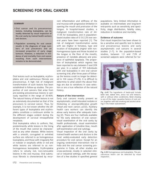
Fig. 4.49 The ingredients of betel-quid include<br />
betel leaf, slaked lime, areca nut and tobacco,<br />
with or without other condiments. Betel chewing<br />
is one of the major causative factors of oral <strong>cancer</strong><br />
(together with bidi smoking and alcohol drinking)<br />
in the Indian subcontinent.<br />
Fig. 4.50 Homogeneous oral leukoplakia. This pre<strong>cancer</strong>ous<br />
lesion can be detected by visual<br />
inspection.


