- Page 1 and 2:
THE UNIVERSITY OF CALGARY Functiona
- Page 3:
Many figures in this thesis use col
- Page 6 and 7:
ACKNOWLEûGEM ENTS Many friends and
- Page 8 and 9:
Approval page Acknowledgements Tabi
- Page 10 and 11:
CONCLUDING COMMENTS Figures for Cha
- Page 12 and 13:
CHAPTER 5: Mechanical and phylogene
- Page 14 and 15:
LIST OF FIGURES Figure 1 .l. Phylog
- Page 16 and 17:
Figure 3.6. Osteological correlates
- Page 18 and 19:
AMNH GI HMN IVPP MOR NMC OMNH PIN P
- Page 20 and 21:
and constructional morphology revea
- Page 22 and 23:
1985, Bryant and Seymour 1990). If
- Page 24 and 25:
surfaces of a Tyrannosaurus rex spe
- Page 26 and 27:
3. Troodontidae (Figure 1.2): Trwdo
- Page 29 and 30:
Hypothesized functions of the tyran
- Page 31 and 32:
1. Atomization (Chapters 2 and 3).
- Page 33 and 34:
Figure 1.1. P hylogenetic diagram o
- Page 35 and 36:
Figure 1.2. Phylogenetic diagram of
- Page 37 and 38:
Figure 1.3. Phylogenetic diagram of
- Page 39 and 40:
Figure 1.4. Phylogenetic diagram of
- Page 41:
Figure 1 .S. Skeletal restorations
- Page 44 and 45:
did not quantify the degree of prox
- Page 46 and 47:
qualitative debate over arctometata
- Page 48 and 49:
This hypothesis focuses on tyrannos
- Page 50 and 51:
Table 2.1. Theropod and Plafeosauru
- Page 52 and 53:
specimens in the figures reflects a
- Page 54 and 55:
were not attempted. While this limi
- Page 56 and 57:
Chapter 3). 1 now address variation
- Page 58 and 59:
- Ç6'P 1 68'€tr 1 L'OC 99'66 L'9
- Page 60 and 61:
Table 2.3: Log-transformed values f
- Page 62 and 63:
experiences a sharp laterally indin
- Page 64 and 65:
ecause it can be cursorily dassifie
- Page 66 and 67:
arctometatarsalian specimens, the l
- Page 68 and 69:
) Shaf€ and region of proximal ar
- Page 70 and 71:
a) Ginglymus. Figure 2.14 shows the
- Page 72 and 73:
) Shaft and region of proximal arti
- Page 74 and 75:
measurements of width pV2S%TU-DE an
- Page 76 and 77:
Table 2.4b Variancelmeasurement eac
- Page 78 and 79:
the largest sum of linear measureme
- Page 80 and 81:
Table 2.5: Specimen statistics Herr
- Page 82 and 83:
and Sinraptor dongi specimens. With
- Page 84 and 85:
In addition, a hook like proximal a
- Page 86 and 87:
oth physical narrowing and elongati
- Page 88 and 89:
1s the MT 111 shape segregation fun
- Page 90 and 91:
tyrannosaurids, ornithomimid, and t
- Page 92 and 93:
proximal constriction, white EImisa
- Page 95:
Figure 2.2. Tyrannosaurid, trwdonti
- Page 99: Figure 2.4. Dromaeosaurid and carno
- Page 102 and 103: - Anterior (flexor) surface medial
- Page 105 and 106: Figure 2.7. Left MT III of Tyrannos
- Page 107 and 108: Figure 2.8. Left MT III of Gorgosau
- Page 109 and 110: Figure 2.9. Left MT III of an omith
- Page 111 and 112: Figure 2.10. Left metatarsus of Tmd
- Page 113 and 114: Figure 2.1 1. Left MT III of Elmisa
- Page 115 and 116: Figure 2.12. Right MT III of Deinon
- Page 117 and 118: Figure 2.13. Left MT III of Allosau
- Page 119 and 120: Figure 2.14. Left metatarsus of Sin
- Page 121 and 122: Figure 2.15. Plots of MT Ill specir
- Page 123: Figure 2.16. Plot of third metatars
- Page 126 and 127: tv . ... . Ri. . . .
- Page 129 and 130: Figure 2.19. Plot of third metatars
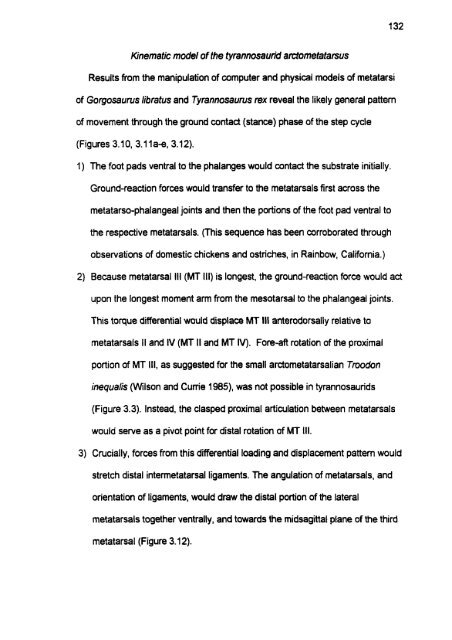
- Page 131 and 132: CHAPTER 3: Tensile keystone model o
- Page 133 and 134: dynamically transmitted Iommotor fo
- Page 135 and 136: Table 3.1 . Metatarsi examined in t
- Page 137 and 138: interrnetatarsal dynamics in these
- Page 139 and 140: compelled analysis of movement evid
- Page 141 and 142: Once prepared, TMP 94.1 2.602 and i
- Page 143 and 144: Daspletosaurus tomsus (MOR 590), an
- Page 145 and 146: 2. Freedom of movement inferred fro
- Page 147 and 148: Table 3.2. Surface areas of intemet
- Page 149: phylogenetic bracketing Wtmer 1995)
- Page 153 and 154: e expected for tyrannosaurid interm
- Page 155 and 156: The preceding discussion derives fr
- Page 157 and 158: weapons sparring), and the arctomet
- Page 159: Figure 3.1. Schematic representatio
- Page 163: Figure 3.3. CT reconstruction of ri
- Page 167: Figure 3.5. MT II (left element) an
- Page 171: Figure 3.7. Osteological correlates
- Page 175: Figure 3.9. Osteological correlates
- Page 179: Figure 3.1 1 ae. The metatarsal rec
- Page 187: Figure 3.13. Ligament contribution
- Page 191 and 192: Figure 3.15. Anatorny of the equid
- Page 193: Figure 3.16. Cornparison of loading
- Page 196 and 197: (nodes), and solving stresslstrain
- Page 198 and 199: The practicality of the finite elem
- Page 200 and 201:
collective density of trabeculae (K
- Page 202 and 203:
A FORCE INPUTS AND MATERIAL PROPERT
- Page 204 and 205:
Alexander et al. (1 979) rewrded a
- Page 206 and 207:
The preceding loading regime entait
- Page 208 and 209:
detemined by using the photographic
- Page 210 and 211:
2. Preparation of data for contour
- Page 212 and 213:
. - Elastic modulus (GP4 Ex EY Ez P
- Page 214 and 215:
conversion. Volume-to-nuages facili
- Page 216 and 217:
maintained. An angle of 2 degrees p
- Page 218 and 219:
of MT III (Figure 4.4, pinpointed b
- Page 220 and 221:
anterodorsal rotation of the distal
- Page 222 and 223:
allowed to stretch slightly under t
- Page 224:
Figure 4.1. Initial loads and bound
- Page 228:
Figure 4.3. Finite element mesh of
- Page 232:
Figure 4.5. Strain distribution in
- Page 236 and 237:
CHAPTER 5: Mechanical and evolution
- Page 238 and 239:
y distal intermetatarsal ligaments,
- Page 240 and 241:
Probable fundion of the tyrannosaur
- Page 242 and 243:
Ornitholestes, and camosaurs) incre
- Page 244 and 245:
morphology matches in degree the co
- Page 246 and 247:
discussion elucidates the evolution
- Page 248 and 249:
face of the skull met part of the m
- Page 250 and 251:
Alternately, a morphological novelt
- Page 252 and 253:
outgroup to ail theropods, and the
- Page 254 and 255:
convincingly evident in figures of
- Page 256 and 257:
parsimonious scenarios are outlined
- Page 258 and 259:
dromaeosaurids], and a dade compris
- Page 260 and 261:
arctometatarsalian forrns. The conv
- Page 262 and 263:
Proximal intermetatarsal ligaments
- Page 264 and 265:
witti a surfeit of potentially supp
- Page 267:
Figure 5.2. Phylogeny of the Therop
- Page 271:
Figure 5.4. Phylogeny of the Therop
- Page 275:
Figure 5.6. Phylogeny of the Therop
- Page 278 and 279:
Bock W.J. and von Wahlert G. Evolut
- Page 280 and 281:
Cume P.J. 1997. Theropoda. In: Fari
- Page 282 and 283:
Grenard S. 1991. Handbook of Alliga
- Page 284 and 285:
Liem K.F. and Osse J.W.M. 1975. Bio
- Page 286 and 287:
Richtsmeier J.T. and Cheverud J.M.
- Page 288 and 289:
Woo S., Maynard J., Butler O. 1987.
- Page 290 and 291:
Different variables in a PCA will h