Analysis of the extended defects in 3C-SiC.pdf - Nelson Mandela ...
Analysis of the extended defects in 3C-SiC.pdf - Nelson Mandela ...
Analysis of the extended defects in 3C-SiC.pdf - Nelson Mandela ...

Create successful ePaper yourself
Turn your PDF publications into a flip-book with our unique Google optimized e-Paper software.
94<br />
left is that <strong>the</strong> l<strong>in</strong>e direction is <strong>of</strong> <strong>the</strong> type. The [100] makes an angle <strong>of</strong> 54.74°<br />
with <strong>the</strong> [111] direction and if one <strong>in</strong>fers <strong>the</strong> foil thickness from <strong>the</strong> projected<br />
dislocation length, a thickness <strong>of</strong> ~280 nm is found. Thus it is possible that <strong>the</strong><br />
dislocation l<strong>in</strong>e direction is <strong>of</strong> <strong>the</strong> 100 type with <strong>the</strong> dislocation not runn<strong>in</strong>g<br />
completely from top to bottom <strong>in</strong> <strong>the</strong> foil but ra<strong>the</strong>r hav<strong>in</strong>g been nucleated at a defect<br />
somewhere with<strong>in</strong> <strong>the</strong> foil. A possible nucleation po<strong>in</strong>t is shown on <strong>the</strong> micrograph <strong>in</strong><br />
Fig. 7.16. by <strong>the</strong> white arrow. Some form <strong>of</strong> residual contrast is visible which may<br />
<strong>in</strong>dicate a defect which acted as nucleation po<strong>in</strong>t.<br />
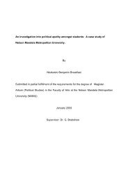
Fig. 7.21. The simulated image <strong>of</strong> <strong>the</strong> dislocation <strong>in</strong>dicated on Fig. 7.16.<br />
None <strong>the</strong> less, <strong>the</strong> identification <strong>of</strong> <strong>the</strong> dislocation was done by simulation and <strong>the</strong><br />
results are shown <strong>in</strong> Fig. 7.21. Excellent agreement is seen but <strong>the</strong> true nature <strong>of</strong> <strong>the</strong><br />
defect will have to be established <strong>in</strong> a future study.<br />
7.4 Reassessment <strong>of</strong> Unimplanted Sample<br />
7.4.1 Overview <strong>of</strong> Investigation<br />
The unimplanted <strong>SiC</strong> sample was <strong>in</strong>vestigated aga<strong>in</strong>, us<strong>in</strong>g a beam direction,<br />
to determ<strong>in</strong>e whe<strong>the</strong>r similar dislocation complexes to those observed <strong>in</strong> <strong>the</strong> hydrogen<br />
implanted <strong>SiC</strong> sample annealed at 1600°C was present. It was found that <strong>the</strong> same<br />
dislocation structures are present <strong>in</strong> <strong>the</strong> unimplanted sample and it was concluded that<br />
dislocation complexes was not <strong>in</strong>troduced as a result <strong>of</strong> <strong>the</strong> irradiation or anneal<strong>in</strong>g<br />
process.