- Page 1 and 2:
I "a f IMods Hole '" ~ o c.,,~tlOGR
- Page 3 and 4:
) )
- Page 5 and 6:
" \ / )
- Page 7 and 8:
parent sediment on the eastern oute
- Page 9 and 10:
esearch. 5 )
- Page 11 and 12:
Res u 1 ts. . . . . . . . . . . . .
- Page 13 and 14:
Figure 3.12 Echo-sounding profile (
- Page 15 and 16:
Fi gure 6.14 6. 15 7. 1 7.2 7.3 7.4
- Page 17 and 18:
\ I ) Table 4. 1 4.2 5.1 5.2 5.3 6.
- Page 19 and 20:
CHAPTER I INTRODUCTION The area inv
- Page 21 and 22:
) \ .1 Fi gure i. 1. Bathymetry of
- Page 23 and 24:
~. ~ 71° 70° tI 68 fJ0 25° 7' 25
- Page 25 and 26:
) ) This is expected since most of
- Page 27:
GENERAL DES CRI PT I ON CHAPTE R I
- Page 30 and 31:
j
- Page 32 and 33:
egi onal contours. An area centered
- Page 34 and 35:
this region. However, all of these
- Page 36 and 37:
) ì /
- Page 38 and 39:
') ) ) /
- Page 41 and 42:
'"-, i~ 7t 70° 6f 68 67° 25° 72
- Page 43:
Fig u r e 3. 2 . CON RA D 10 s e is
- Page 46 and 47:
sequence corresponds to layered tur
- Page 49:
) . .) Figure 3.3. Map showing domi
- Page 52 and 53:
\ )
- Page 55 and 56:
"- '''--.- 25° ................. .
- Page 57 and 58:
) ) others, 1973), and the upper po
- Page 59 and 60:
) Figure 3.5. CONRAD 8 seismic refl
- Page 61 and 62:
. Q) 3W/L NOI.L~ l( . (Y CI s. :: 0
- Page 63 and 64:
.) Figure 3.6. Idealized profile al
- Page 65 and 66:
) ) a: 0 ~ ~ (J L. Z Q) a: ~ lU UJ
- Page 67 and 68:
) ) Figure 3.7. CONRAD 10 seismic r
- Page 69 and 70:
i- (Y (1 ~:: en or- LL 47
- Page 71 and 72:
) ) THE TRANSPARENT LAYER The distr
- Page 73 and 74:
Fi gure 3.8. CH A I N 34 s e ism i
- Page 75 and 76:
co . (Y (l So :: O' "r- Li 51
- Page 77 and 78:
) 53
- Page 79 and 80:
) Figure 3.9. CONRAD 10 seismic ref
- Page 81 and 82:
0' (V CJ s. :: O' ... LL 55
- Page 83 and 84:
\ J ) Figure 3.10. Seismic reflecti
- Page 85 and 86:
or- . (V Q) S- :: rn Li 58
- Page 87 and 88:
ì ) Figure 3.11. CONRAD 10 seismic
- Page 89 and 90:
l UJ .líliiiP',,:... ¡ '"'" ~ ' ~
- Page 91 and 92:
) .I Figure 3.12. Echo-soundi ng pr
- Page 93 and 94:
N CV OJ s. :: 01 l. 62
- Page 95 and 96:
) ) Figure 3.13. CONRAD 10 seismic
- Page 97 and 98:
(' r- (' (j $. :: en 'r- LL 64
- Page 99 and 100:
) ) GENERAL LITHOLOGY CHAPTER iv TH
- Page 101 and 102:
~ , ) Figure 4.1. General lithology
- Page 103:
.'-/ '..-/ AIr 60-8 A II 60-8 An60~
- Page 106 and 107:
) )
- Page 108 and 109:
average of 80% lutite (~ 2 ~m) and
- Page 110 and 111:
) )
- Page 112 and 113:
.\ ! ') )
- Page 114 and 115:
ì/ )
- Page 116 and 117:
10 m2, with an average value of 1.3
- Page 118 and 119:
) ) /
- Page 120 and 121:
The laminated zones and overlying p
- Page 122 and 123:
) )
- Page 124 and 125:
\ j )
- Page 127 and 128:
) ) Figure 4.7. Graph of carbonate
- Page 129 and 130:
) ) z 0 N O~ E I i ,,_.' i." I ." .
- Page 131:
) ì i Figure 4.8. Curves of carbon
- Page 134 and 135:
currents have transported many Fora
- Page 136 and 137:
increased dissolution (elevated lys
- Page 138 and 139:
TABLE 4.1. MINERALOGY OF THE c 2 ~m
- Page 140 and 141:
the following peaks and weighting f
- Page 142 and 143:
.,1 ì / ) j
- Page 144 and 145:
\ J )
- Page 146 and 147:
) )
- Page 148 and 149:
) )
- Page 150 and 151:
Obvi ous ly, di 1 uti on of the nor
- Page 152 and 153:
The m i n era log i c a 1 com po si
- Page 154 and 155:
gray 1 uti t e s per s i s t n ear
- Page 156 and 157:
) )
- Page 158 and 159:
TABLE 5.1. AVERAGE RATES OF SEDIMEN
- Page 160 and 161:
and thus the T-Z zonation (Fig. 5.1
- Page 162 and 163:
) ) )
- Page 164 and 165:
TABLE 5.2. AVERAGE RATES OF SEDIMEN
- Page 166 and 167:
Cl z: c: l/ l. i- c: Cl z: o OJ IX
- Page 168 and 169:
consolidated (A.J. Silva, pers. com
- Page 170 and 171:
) ) _/
- Page 172 and 173:
\ ) \ ì ../
- Page 174 and 175:
in age, and the ash is probably ass
- Page 176 and 177:
') .I \...../
- Page 178 and 179:
122 must result from mixing with th
- Page 180 and 181:
eas twa rd flow of NADW between 150
- Page 182 and 183:
whi ch recorded currents for about
- Page 184 and 185:
,) , ,
- Page 186 and 187:
ottom photographs in February, 1972
- Page 188 and 189:
Several important features were obs
- Page 190 and 191:
. \ .' ì ,) ~)
- Page 192 and 193:
. ) )
- Page 194 and 195:
)j )
- Page 196 and 197:
) )
- Page 198 and 199:
, " ) ) ./
- Page 200 and 201:
. \ ) )
- Page 202 and 203:
'\ ) ) J
- Page 204 and 205:
I )
- Page 206 and 207:
141 turn north in the western part
- Page 208 and 209:
" ) , \ -./)
- Page 210 and 211:
. ) ) / )
- Page 212 and 213:
145 bottle spacing. An example is t
- Page 215 and 216:
~() ~ 3.0 ~ ~ ) ~ ~i. ) .. ~ 2.0 ~i
- Page 217 and 218:
) ) Figure 6.6. Silicate ("g-atoms/
- Page 219 and 220:
) ) ~ 2.2 ~ 2.0 t2 ~ tt ~" 1.8 .. ~
- Page 221 and 222:
,) -) 152 structure persist at and
- Page 223 and 224:
) ~) 154 Figure 6.7. Current patter
- Page 225 and 226:
) ) 72° DEPTH IN CORR. METERS Fi g
- Page 227 and 228:
) ) speeds of 2 cm/sec (the stall s
- Page 229 and 230:
) I / Fi gure6. 8. 158 Progressi ve
- Page 231 and 232:
" ) / ) 10 19 17 5 5616 M 5801 M N
- Page 233:
) ) ~ 160 Figure 6.9. Polar histogr
- Page 236 and 237:
. ì )
- Page 239 and 240:
,) B(LOWER) START 22 B(UPPER) START
- Page 241 and 242:
) ) Figure 6.11. Polar histogram of
- Page 243 and 244:
) 2700 ) 00 1800 F; g u re 6. 11 C
- Page 245 and 246:
, ) ) _/ Figure 6.12. 168 Progress
- Page 247 and 248:
\ --~ E START 21 5 30 12 XI 21 xu )
- Page 249 and 250:
) / i I .~J the flow recorded at cu
- Page 251 and 252: ) Figure 6.13. Directions and relat
- Page 253 and 254: ') ./ ) ,j 70° 68° 66° ~~oo · "
- Page 255: Fig u re 6. 14. i 75 Bottom photogr
- Page 258 and 259: S e c t ion 2 (F i g s. 6. 1, 6. 7)
- Page 260 and 261: ) )
- Page 262 and 263: ') / ) j
- Page 264 and 265: northwest flow along the west flank
- Page 266 and 267: " ) /
- Page 268 and 269: \ )
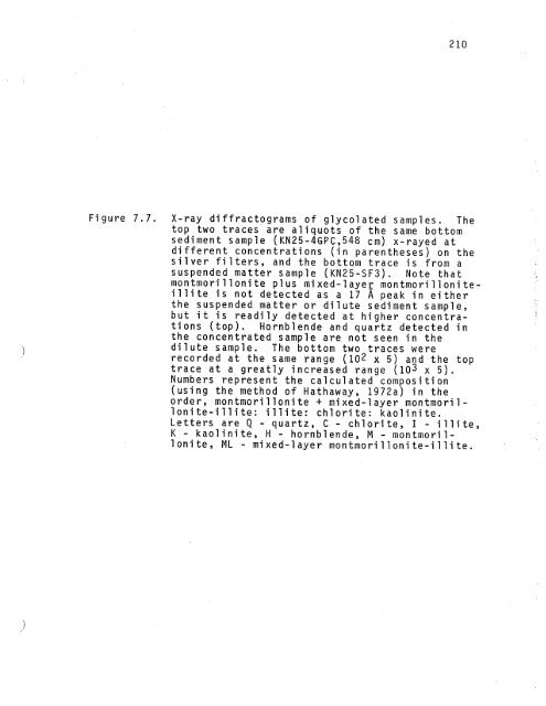
- Page 270 and 271: filter (0.50 lJmpore size). After f
- Page 273 and 274: . '! ) ) ,./ 2.2 ~ 2.i () '- 2.0 ~
- Page 275 and 276: ) ) Figure 7.3. Light-scattering pr
- Page 277 and 278: '\. './. V 23-7 V 23-6 V 25-74 V 25
- Page 279 and 280: extensive erosion on the lower cont
- Page 281 and 282: ) \/ (SEM) to determine composition
- Page 283: \ ) ) Figure 7.4. 195 Scanning elec
- Page 286 and 287: ) \ ./
- Page 288 and 289: 'i j
- Page 290 and 291: The less than 2 ~m (lutite) fractio
- Page 292 and 293: 201 In a sample where additional cl
- Page 294 and 295: TAB LE 7. 1. CALCULATE D CLAY MI NE
- Page 296 and 297: :1 / , ì j
- Page 298 and 299: I ) \ )
- Page 300 and 301: cannot always be accepted at face v
- Page 304 and 305: , \! )
- Page 306 and 307: in favor of the concept that the su
- Page 308 and 309: on the north flank of the far easte
- Page 310 and 311: that enough suspended matter has be
- Page 312 and 313: The following pages describe a mode
- Page 314 and 315: ) )
- Page 316 and 317: series of ridges associated with an
- Page 318 and 319: Eocene to 01 i gocene (Monroe, 1968
- Page 320 and 321: :') ¡i )
- Page 322 and 323: \ i / ') ./
- Page 324 and 325: (Table 8.1). The interbedded silts
- Page 326 and 327: 229 Chase and Hersey, 1968). The th
- Page 329 and 330: \ ! Figure 8.3. 231 Schematic diagr
- Page 332 and 333: Downslope sedimentation covered the
- Page 335 and 336: ) 235 Fi gure 8.4. Sketch showi ng
- Page 337 and 338: '\ l ) ~z w u w a: o ~ wz w o u ~ w
- Page 339 and 340: ) / the same event of current-contr
- Page 341 and 342: ) Figure 8.5. Idealized representat
- Page 343 and 344: ) \ ) d / 'I i II I Ii I i I i I I
- Page 345 and 346: ) \ ) formed earlier depositional s
- Page 347 and 348: ) ) homogenei ty of the sediments o
- Page 349: ) ) 2 ) Sou t h S lop e - P u e rt
- Page 352 and 353:
248 Biscaye, P.E. and S.L. Eittreim
- Page 354 and 355:
Ericson, D.B., M. Ewing, G. Wollin
- Page 356 and 357:
Hersey, J.B. (1965) Sediment pondin
- Page 358 and 359:
McCullough, J.R. and G.H. Tupper (1
- Page 360 and 361:
Swallow, J.C. and L.V. Worthington
- Page 362 and 363:
" ) )
- Page 364 and 365:
259 approximation of the amount of
- Page 366 and 367:
s ~ 0 ~ Q) o '- CO 0 U ~ it \f0 Q)
- Page 368 and 369:
) )
- Page 370 and 371:
Crui se All 11 All 11 AII 60 Leg 8
- Page 372 and 373:
TABLE A2.1. BOTTOM PHOTOGRAPHS (Con
- Page 374 and 375:
TABLE A2.1. BOTTOM PHOTOGRAPHS (Con
- Page 376 and 377:
TAB LE A2. 1. BOTTOM PHOTOGRAPHS (C
- Page 378 and 379:
CH 19 CH 19 CH 19 CH 34 CH 34 TABLE
- Page 380 and 381:
KN25 KN 25 KN 25 KN 25 TABLE A2.1.
- Page 382 and 383:
LL 8 LL 8 LL 8 LL 8 LL 8 SA C ru is
- Page 384 and 385:
) )
- Page 386 and 387:
s. .. M i. 0 1. " .. QJ ..- N .. C'
- Page 388 and 389:
~.. c. 0' 0 .. Q)+l- ~ 0' 0' 0 ~ M
- Page 390 and 391:
TABLE A3.1. SUSPENDED PARTICULATE M
- Page 393 and 394:
) -) APPENDIX IV PHYSICAL PROPERTIE
- Page 395:
) ) Figure A4.1. Plots of water con
- Page 398 and 399:
) \ )
- Page 400 and 401:
ì )
- Page 402 and 403:
\ )
- Page 404 and 405:
') /
- Page 406 and 407:
\ ) ) . //
- Page 409 and 410:
) ) C\' 0 § to ~ ~ ~ 0 ~ ~ ~ CI Q:
- Page 411 and 412:
) /) 295 Figure A4.5. Plots of wate
- Page 413 and 414:
'-. ~, 160 SHEAR STRENGTH 30 50 (g/
- Page 415:
) ) Figure A4.6. Plots of water con
- Page 418 and 419:
) . ) .
- Page 420 and 421:
ì ) )
- Page 422 and 423:
\ ) )
- Page 424 and 425:
.J , .)
- Page 426 and 427:
ì /"
- Page 428 and 429:
D \ )
- Page 430 and 431:
\ / ) /
- Page 432 and 433:
) , )
- Page 435 and 436:
) ) 307 Fi gure A4.11. Plots of wat
- Page 437 and 438:
"-, ',, WATER CONTENT (% DRY WEIGHT
- Page 439:
) j 309 Figure A4.12. Plots of wate
- Page 442 and 443:
) ) ./
- Page 445 and 446:
) 312 WATER CONTENT (%DRY WT) SHEAR
- Page 447 and 448:
) J BIOGRAPHY Born: Ma rch 19, 1946
- Page 449:
') \ ) ~UWDATORY DISTRIBUTION LIST
- Page 452 and 453:
) J
- Page 454:
UNCLASSI FIED 1/31/74 SECURITY CLAS