

Low_resolution_Thesis_CDD_221009_public - Visual Optics and ...
Low_resolution_Thesis_CDD_221009_public - Visual Optics and ...
Low_resolution_Thesis_CDD_221009_public - Visual Optics and ...

Create successful ePaper yourself
Turn your PDF publications into a flip-book with our unique Google optimized e-Paper software.
REFRACTIVE SURGERY PMMA MODEL<br />
3<br />
2<br />
Measured spheres<br />
With experimental efficency factor<br />
With theoretical efficency factor<br />
No efficiency effects (fat surfaces)<br />
Asphericity<br />
1<br />
0<br />
-1<br />
0 3 6 9 12 15<br />
Correction (D)<br />
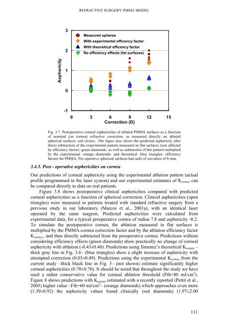
Fig. 3.7. Postoperative corneal asphericities of ablated PMMA surfaces as a function<br />
of nominal (on cornea) refractive correction, as measured directly on ablated<br />
spherical surfaces -red circles-. The figure also shows the predicted asphericity after<br />
direct subtraction of the experimental pattern measured on flat surfaces (non affected<br />
by efficiency factor) -green diamonds- as well as subtraction of this pattern multiplied<br />
by the experimental -orange diamonds- <strong>and</strong> theoretical -blue triangles- efficiency<br />
factors for PMMA. Pre-operative spherical surfaces had radii of curvature of 8-mm.<br />
3.4.5. Post - operative asphericities on cornea<br />
Our predictions of corneal asphericity using the experimental ablation pattern (actual<br />
profile programmed in the laser system) <strong>and</strong> our experimental estimates of K cornea can<br />
be compared directly to data on real patients.<br />
Figure 3.8 shows postoperative clinical asphericities compared with predicted<br />
corneal asphericities as a function of spherical correction. Clinical asphericities (open<br />
triangles) were measured in patients treated with st<strong>and</strong>ard refractive surgery from a<br />
previous study in our laboratory (Marcos et al., 2001a), with an identical laser<br />
operated by the same surgeon. Predicted asphericities were calculated from<br />
experimental data, for a typical preoperative cornea of radius 7.8 <strong>and</strong> asphericity -0.2.<br />
To simulate the postoperative cornea, the ablation measured in flat surfaces is<br />
multiplied by the PMMA-cornea correction factor <strong>and</strong> by the ablation efficiency factor<br />
K cornea , <strong>and</strong> then directly subtracted from the preoperative cornea. Predictions without<br />
considering efficiency effects (green diamonds) show practically no change of corneal<br />
asphericity with ablation (-0.43±0.40). Predictions using Jimenez’s theoretical K cornea –<br />
thick gray line in Fig. 3.6– (blue triangles) show a slight increase of asphericity with<br />
attempted correction (0.03±0.49). Predictions using the experimental K cornea from the<br />
current study –thick black line in Fig. 3– (not shown) estimate significantly higher<br />
corneal asphericities (0.78±0.70). It should be noted that throughout the study we have<br />
used a rather conservative value for corneal ablation threshold (Fth=40 mJ/cm 2 ).<br />
Figure 8 shows predictions with K cornea estimated with a recently reported (Pettit et al.,<br />
2005) higher value –Fth=60 mJ/cm 2 – (orange diamonds) which approaches even more<br />
(1.39±0.92) the asphericity values found clinically (red diamonds) (1.97±2.00<br />
111


