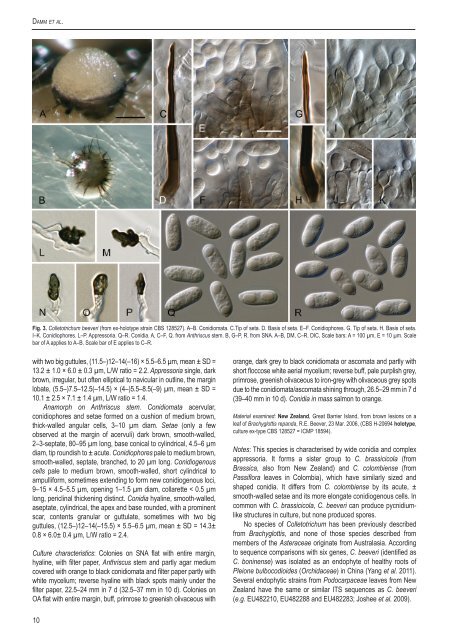
Damm et al. Fig. 3. <strong>Colletotrichum</strong> beeveri (from ex-holotype strain <strong>CBS</strong> 128527). A–B. Conidiomata. C.Tip of seta. D. Basis of seta. E–F. Conidioph<strong>or</strong>es. G. Tip of seta. H. Basis of seta. I–K. Conidioph<strong>or</strong>es. L–P. Appress<strong>or</strong>ia. Q–R. Conidia. A, C–F, Q. from Anthriscus stem. B, G–P, R. from SNA. A–B, DM, C–R. DIC, Scale bars: A = 100 µm, E = 10 µm. Scale bar of A applies to A–B. Scale bar of E applies to C–R. with two big guttules, (11.5–)12–14(–16) × 5.5–6.5 µm, mean ± SD = 13.2 ± 1.0 × 6.0 ± 0.3 µm, L/W ratio = 2.2. Appress<strong>or</strong>ia single, dark brown, irregular, but often elliptical to navicular in outline, the margin lobate, (5.5–)7.5–12.5(–14.5) × (4–)5.5–8.5(–9) µm, mean ± SD = 10.1 ± 2.5 × 7.1 ± 1.4 µm, L/W ratio = 1.4. Anam<strong>or</strong>ph on Anthriscus stem. Conidiomata acervular, conidioph<strong>or</strong>es and setae f<strong>or</strong>med on a cushion of medium brown, thick-walled angular cells, 3–10 µm diam. Setae (only a few observed at the margin of acervuli) dark brown, smooth-walled, 2–3-septate, 80–95 µm long, base conical to cylindrical, 4.5–6 µm diam, tip roundish to ± acute. Conidioph<strong>or</strong>es pale to medium brown, smooth-walled, septate, branched, to 20 µm long. Conidiogenous cells pale to medium brown, smooth-walled, sh<strong>or</strong>t cylindrical to ampullif<strong>or</strong>m, sometimes extending to f<strong>or</strong>m new conidiogenous loci, 9–15 × 4.5–5.5 µm, opening 1–1.5 µm diam, collarette < 0.5 µm long, periclinal thickening distinct. Conidia hyaline, smooth-walled, aseptate, cylindrical, the apex and base rounded, with a prominent scar, contents granular <strong>or</strong> guttulate, sometimes with two big guttules, (12.5–)12–14(–15.5) × 5.5–6.5 µm, mean ± SD = 14.3± 0.8 × 6.0± 0.4 µm, L/W ratio = 2.4. Culture characteristics: Colonies on SNA flat with entire margin, hyaline, with filter paper, Anthriscus stem and partly agar medium covered with <strong>or</strong>ange to black conidiomata and filter paper partly with white mycelium; reverse hyaline with black spots mainly under the filter paper, 22.5–24 mm in 7 d (32.5–37 mm in 10 d). Colonies on OA flat with entire margin, buff, primrose to greenish olivaceous with <strong>or</strong>ange, dark grey to black conidiomata <strong>or</strong> ascomata and partly with sh<strong>or</strong>t floccose white aerial mycelium; reverse buff, pale purplish grey, primrose, greenish olivaceous to iron-grey with olivaceous grey spots due to the conidiomata/ascomata shining through, 26.5–29 mm in 7 d (39–40 mm in 10 d). Conidia in mass salmon to <strong>or</strong>ange. Material examined: New Zealand, Great Barrier Island, from brown lesions on a leaf of Brachyglottis repanda, R.E. Beever, 23 Mar. 2006, (<strong>CBS</strong> H-20694 holotype, culture ex-type <strong>CBS</strong> 128527 = ICMP 18594). Notes: This <strong>species</strong> is characterised by wide conidia and <strong>complex</strong> appress<strong>or</strong>ia. It f<strong>or</strong>ms a sister group to C. brassicicola (from Brassica, also from New Zealand) and C. colombiense (from Passifl<strong>or</strong>a leaves in Colombia), which have similarly sized and shaped conidia. It differs from C. colombiense by its acute, ± smooth-walled setae and its m<strong>or</strong>e elongate conidiogenous cells. In common with C. brassicicola, C. beeveri can produce pycnidiumlike structures in culture, but none produced sp<strong>or</strong>es. No <strong>species</strong> of <strong>Colletotrichum</strong> has been previously described from Brachyglottis, and none of those <strong>species</strong> described from members of the Asteraceae <strong>or</strong>iginate from Australasia. Acc<strong>or</strong>ding to sequence comparisons with six genes, C. beeveri (identified as C. boninense) was isolated as an endophyte of healthy roots of Pleione bulbocodioides (Orchidaceae) in China (Yang et al. 2011). Several endophytic strains from Podocarpaceae leaves from New Zealand have the same <strong>or</strong> similar ITS sequences as C. beeveri (e.g. EU482210, EU482288 and EU482283; Joshee et al. 2009). 10
The <strong>Colletotrichum</strong> boninense <strong>species</strong> <strong>complex</strong> <strong>Colletotrichum</strong> boninense M<strong>or</strong>iwaki, Toy. Sato & Tsukib., Mycoscience 44(1): 48. 2003. Fig. 4. Teleom<strong>or</strong>ph developed on OA (<strong>CBS</strong> 123756). Ascomata perithecia, variable in shape but usually subglobose to pyrif<strong>or</strong>m, glabrous, medium brown, 100–300 × 100–200 µm, ostiolate, periphysate, neck hyaline to pale brown, to 100 µm in length, outer wall composed of flattened angular cells 4–15 µm diam. Interascal tissue composed of rather irregular thin-walled hyaline septate paraphyses. Asci in a basal fascicle, cylindric-clavate, 45–60 × 12.5–17 µm, 8-sp<strong>or</strong>ed, with a ± truncate apex and a small refractive apical ring. Ascosp<strong>or</strong>es initially hyaline and aseptate, becoming 1–3-septate, septation sometimes occurring inside the ascus, light to medium brown-pigmented, sometimes verruculose pri<strong>or</strong> to the start of germination, allantoid, (12.5–)14–17(–18) × (4–)5–6(–6.5) µm, mean ± SD = 15.6 ± 1.4 × 5.4 ± 0.5 µm, L/W ratio = 2.9. Anam<strong>or</strong>ph developed on SNA (<strong>CBS</strong> 123755). Vegetative hyphae 1–6 µm diam, hyaline <strong>or</strong> pale brown, smooth-walled, septate, branched. Chlamydosp<strong>or</strong>es not observed. Conidiomata po<strong>or</strong>ly <strong>or</strong> not developed, conidioph<strong>or</strong>es and setae f<strong>or</strong>med directly on hyphae. Setae rare, medium brown, smooth to verruculose, 1–2-septate, 20–60 µm long, base cylindrical, conical <strong>or</strong> slightly inflated, 3–7 µm diam at the widest part, tip ± rounded. Conidioph<strong>or</strong>es hyaline <strong>or</strong> pale brown, simple <strong>or</strong> septate, branched <strong>or</strong> unbranched, to 40 µm long. Conidiogenous cells hyaline <strong>or</strong> pale brown, cylindrical, 6–15 × 3–5 µm, opening 1–2 µm diam, collarette 0.5–1.5 µm long, periclinal thickening conspicuous. Conidia hyaline, smoothwalled, aseptate, straight, cylindrical, apex round, base round with a prominent hilum, often containing two big polar guttules, (8.5–)11–14.5(–17.5) × (4–)5–6(–6.5) µm, mean ± SD = 12.8 ± 1.6 × 5.4 ± 0.4 µm, L/W ratio = 2.4. Appress<strong>or</strong>ia solitary <strong>or</strong> in sh<strong>or</strong>t chains, medium brown, thick-walled, entire edge <strong>or</strong> crenate, rarely lobate, smooth-walled, irregular in shape, but often bullet-shaped <strong>or</strong> navicular with an acute tip, (4.5–)7–14(–18) × (4–)5–8(–11) µm, mean ± SD = 10.5 ± 3.3 × 6.4 ± 1.5 µm, L/W ratio = 1.6. Anam<strong>or</strong>ph developed on Anthriscus stem (<strong>CBS</strong> 123755). Conidiomata acervular, conidioph<strong>or</strong>es and setae f<strong>or</strong>med from a cushion of pale brown, roundish to angular cells, 3–9 µm diam. Setae rare, medium brown, basal cell often paler, verruculose, 1–2-septate, 30–70 µm long, base cylindrical, conical <strong>or</strong> slightly inflated, 3.5–6.5 µm diam, tip ± round to ± acute. Conidioph<strong>or</strong>es pale brown, septate, branched <strong>or</strong> unbranched, to 40 µm long. Conidiogenous cells pale brown, cylindrical to ellipsoidal, 5.8–17 × 3.5–6 µm, opening 0.5–1.5 µm diam, collarette ≤ 0.5 µm long, periclinal thickening visible to conspicuous. Conidia hyaline, smooth-walled, aseptate, straight, cylindrical to clavate, apex round, base round with a prominent hilum, sometimes with two big polar guttules, (9–)12–14.5(–16.5) × (4–)5.5–6.5 µm, mean ± SD = 13.2 ± 1.4 × 5.8 ± 0.5 µm, L/W ratio = 2.3. The conidia of <strong>CBS</strong> 129831 are longer (up to 20 µm) with an average L/W ratio of 2.6. Culture characteristics: Colonies on SNA flat with slightly undulate margin, hyaline with felty white aerial mycelium on filter paper; reverse filter paper partly pale cinnamon to pale hazel; 25.5–29 mm in 7 d (37.5–40 mm in 10 d). Colonies on OA flat with entire margin; surface covered with felty white, rosy buff <strong>or</strong> very pale glaucous grey aerial mycelium, in the centre pale luteous aerial mycelium; reverse buff, rosy buff, pale luteous to honey-coloured; 27.5–32.5 mm in 7 d (39–40 mm in 10 d). Conidia in mass salmon. <strong>CBS</strong> 102667 is slower growing: SNA 18–21 mm in 7 d (29–29.5 mm in 10 d), OA 21.3–22.5 mm in 7 d (31.5–32.5 mm in 10 d). Material examined: Japan, Bonin Islands, from a diseased leaf of Crinum asiaticum var. sinicum, 1988, T. Sato, culture ex-holotype <strong>CBS</strong> 123755 = MAFF 305972; Bonin Islands, from Crinum asiaticum var. sinicum, 1990, T. Sato, culture <strong>CBS</strong> 123756 = MAFF 306094. Australia, from Leucospermum sp., culture <strong>CBS</strong> 129831 = STE-U 2965. New Zealand, N<strong>or</strong>thland, Kaipara, from flowers of Solanum betaceum, 1 Feb. 2004, M. Manning, culture <strong>CBS</strong> 128549 = ICMP 15444. Notes: Conidia of C. boninense are similar to those of C. karstii, although the ascosp<strong>or</strong>es of C. boninense are m<strong>or</strong>e unif<strong>or</strong>m with rounded ends, becoming brown and septate with age and the asci are longer and wider. We recognise that there is significant genetic variation in C. boninense. Host plants of C. boninense s. str. are very diverse including Amaryllidaceae, Bignoniaceae, Podocarpaceae, Proteaceae, Solanaceae and Theaceae. Several ITS sequences, f<strong>or</strong> example HM044131 (Yuan et al., unpubl. data) from Oryza granulata, and FJ449913 (Hu & Guo, unpubl. data) from Dendrobium sp., both presumably from China, are similar to the ITS of C. boninense, C. oncidii and C. cymbidiicola, but these <strong>species</strong> can not be separated from each other by comparison of ITS sequences. <strong>Colletotrichum</strong> brasiliense Damm, P.F. Cannon, Crous & Massola, sp. nov. MycoBank MB560736. Fig. 5. Etymology: Named after the country where it was collected, Brazil. Teleom<strong>or</strong>ph not observed. Anam<strong>or</strong>ph on SNA. Vegetative hyphae 1–5.5 µm diam, hyaline, smooth-walled, septate, branched. Chlamydosp<strong>or</strong>es not observed. Conidiomata acervular, conidioph<strong>or</strong>es and setae f<strong>or</strong>med on a cushion of pale brown, ± thinwalled, angular cells 3–9 µm diam, however, in strain <strong>CBS</strong> 128528 conidioph<strong>or</strong>es and setae are f<strong>or</strong>med directly on hyphae. Setae sparse, pale to medium brown, basal cell usually paler, smooth to finely verruculose, 2–4-septate, 50–60 µm long, base cylindrical to conical, 6–8 µm diam, tip ± acute to slightly roundish <strong>or</strong> zigzag-shaped. Conidioph<strong>or</strong>es hyaline to pale brown, smooth-walled, simple <strong>or</strong> septate and branched, to 30 µm long. Conidiogenous cells hyaline to pale brown, smooth-walled, cylindrical to ellipsoidal, encased in a mucous sheath, sometimes extending to f<strong>or</strong>m new conidiogenous loci, 7–14 × 4.5–7.5 µm, opening 1–2 µm diam, collarette visible, ≤ 0.5 µm long, periclinal thickening visible, in strain <strong>CBS</strong> 128528 conidiogenous cells longer (12–25 µm) and periclinal thickening conspicuous. Conidia hyaline, smooth-walled, aseptate, cylindrical, the apex and base rounded, with a prominent scar, contents granular <strong>or</strong> guttulate, (11.5–)13–16(–18) × 5–5.5(–6) µm, mean ± SD = 14.6 ± 1.6 × 5.4 ± 0.2 µm, L/W ratio = 2.7, conidia of strain <strong>CBS</strong> 128528 longer, measuring (13.5–)14–19(– 22.5) × (4.5–)5–5.5(–6) µm, mean ± SD = 16.5 ± 2.4 × 5.3 ± 0.3 µm, L/W ratio = 3.1. Appress<strong>or</strong>ia medium to dark brown, smoothwalled, lobed, often with a roundish outline, sometimes also triangular, SNA (5.5–)7–16(–32) × (4–)6.5–13(–24) µm, mean ± SD = 11.5 ± 4.5 × 9.7 ± 3.3 µm, L/W ratio = 1.2. Anam<strong>or</strong>ph on Anthriscus stem. Conidiomata acervular, conidioph<strong>or</strong>es and setae f<strong>or</strong>med on a cushion of pale brown, angular cells, 3–8 µm diam. Setae (only one observed) medium brown, smooth-walled, 3-septate, 65 µm long, base cylindrical, 4.5 µm diam, tip ± acute and zig-zag-shaped. Conidioph<strong>or</strong>es hyaline to pale brown, smooth-walled, simple <strong>or</strong> septate and branched, to 20 µm long. Conidiogenous cells hyaline to pale brown, smoothwalled, cylindrical to ellipsoidal, sometimes extending to f<strong>or</strong>m new conidiogenous loci, 6–12 × 3.5–7.5 µm, opening 1–2 µm diam, collarette 1 µm long, periclinal thickening visible, in strain <strong>CBS</strong> www.studiesinmycology.<strong>or</strong>g 11
- Page 1: Studies in Mycology 73 (September 2
- Page 4 and 5: Studies in Mycology The Studies in
- Page 6 and 7: DEDICATION This volume is dedicated
- Page 9 and 10: available online at www.studiesinmy
- Page 11 and 12: The Colletotrichum boninense specie
- Page 13 and 14: The Colletotrichum boninense specie
- Page 15 and 16: The Colletotrichum boninense specie
- Page 17: The Colletotrichum boninense specie
- Page 21 and 22: The Colletotrichum boninense specie
- Page 23 and 24: The Colletotrichum boninense specie
- Page 25 and 26: The Colletotrichum boninense specie
- Page 27 and 28: The Colletotrichum boninense specie
- Page 29 and 30: The Colletotrichum boninense specie
- Page 31 and 32: The Colletotrichum boninense specie
- Page 33 and 34: The Colletotrichum boninense specie
- Page 35 and 36: The Colletotrichum boninense specie
- Page 37 and 38: The Colletotrichum boninense specie
- Page 39 and 40: The Colletotrichum boninense specie
- Page 41 and 42: The Colletotrichum boninense specie
- Page 43 and 44: The Colletotrichum boninense specie
- Page 45 and 46: available online at www.studiesinmy
- Page 47 and 48: The Colletotrichum acutatum species
- Page 49 and 50: The Colletotrichum acutatum species
- Page 51 and 52: The Colletotrichum acutatum species
- Page 53 and 54: The Colletotrichum acutatum species
- Page 55 and 56: The Colletotrichum acutatum species
- Page 57 and 58: The Colletotrichum acutatum species
- Page 59 and 60: The Colletotrichum acutatum species
- Page 61 and 62: The Colletotrichum acutatum species
- Page 63 and 64: The Colletotrichum acutatum species
- Page 65 and 66: The Colletotrichum acutatum species
- Page 67 and 68: The Colletotrichum acutatum species
- Page 69 and 70:
The Colletotrichum acutatum species
- Page 71 and 72:
The Colletotrichum acutatum species
- Page 73 and 74:
The Colletotrichum acutatum species
- Page 75 and 76:
The Colletotrichum acutatum species
- Page 77 and 78:
The Colletotrichum acutatum species
- Page 79 and 80:
The Colletotrichum acutatum species
- Page 81 and 82:
The Colletotrichum acutatum species
- Page 83 and 84:
The Colletotrichum acutatum species
- Page 85 and 86:
The Colletotrichum acutatum species
- Page 87 and 88:
The Colletotrichum acutatum species
- Page 89 and 90:
The Colletotrichum acutatum species
- Page 91 and 92:
The Colletotrichum acutatum species
- Page 93 and 94:
The Colletotrichum acutatum species
- Page 95 and 96:
The Colletotrichum acutatum species
- Page 97 and 98:
The Colletotrichum acutatum species
- Page 99 and 100:
The Colletotrichum acutatum species
- Page 101 and 102:
The Colletotrichum acutatum species
- Page 103 and 104:
The Colletotrichum acutatum species
- Page 105 and 106:
The Colletotrichum acutatum species
- Page 107 and 108:
The Colletotrichum acutatum species
- Page 109 and 110:
The Colletotrichum acutatum species
- Page 111 and 112:
The Colletotrichum acutatum species
- Page 113 and 114:
The Colletotrichum acutatum species
- Page 115 and 116:
The Colletotrichum acutatum species
- Page 117 and 118:
The Colletotrichum acutatum species
- Page 119 and 120:
The Colletotrichum acutatum species
- Page 121 and 122:
The Colletotrichum acutatum species
- Page 123 and 124:
available online at www.studiesinmy
- Page 125 and 126:
The Colletotrichum gloeosporioides
- Page 127 and 128:
The Colletotrichum gloeosporioides
- Page 129 and 130:
The Colletotrichum gloeosporioides
- Page 131 and 132:
The Colletotrichum gloeosporioides
- Page 133 and 134:
The Colletotrichum gloeosporioides
- Page 135 and 136:
The Colletotrichum gloeosporioides
- Page 137 and 138:
The Colletotrichum gloeosporioides
- Page 139 and 140:
The Colletotrichum gloeosporioides
- Page 141 and 142:
The Colletotrichum gloeosporioides
- Page 143 and 144:
The Colletotrichum gloeosporioides
- Page 145 and 146:
The Colletotrichum gloeosporioides
- Page 147 and 148:
The Colletotrichum gloeosporioides
- Page 149 and 150:
The Colletotrichum gloeosporioides
- Page 151 and 152:
The Colletotrichum gloeosporioides
- Page 153 and 154:
The Colletotrichum gloeosporioides
- Page 155 and 156:
The Colletotrichum gloeosporioides
- Page 157 and 158:
The Colletotrichum gloeosporioides
- Page 159 and 160:
The Colletotrichum gloeosporioides
- Page 161 and 162:
The Colletotrichum gloeosporioides
- Page 163 and 164:
The Colletotrichum gloeosporioides
- Page 165 and 166:
The Colletotrichum gloeosporioides
- Page 167 and 168:
The Colletotrichum gloeosporioides
- Page 169 and 170:
The Colletotrichum gloeosporioides
- Page 171 and 172:
The Colletotrichum gloeosporioides
- Page 173 and 174:
The Colletotrichum gloeosporioides
- Page 175 and 176:
The Colletotrichum gloeosporioides
- Page 177 and 178:
The Colletotrichum gloeosporioides
- Page 179 and 180:
The Colletotrichum gloeosporioides
- Page 181 and 182:
The Colletotrichum gloeosporioides
- Page 183 and 184:
The Colletotrichum gloeosporioides
- Page 185 and 186:
The Colletotrichum gloeosporioides
- Page 187 and 188:
The Colletotrichum gloeosporioides
- Page 189 and 190:
available online at www.studiesinmy
- Page 191 and 192:
Colletotrichum - current status and
- Page 193 and 194:
Colletotrichum - current status and
- Page 195 and 196:
Colletotrichum - current status and
- Page 197 and 198:
Colletotrichum - current status and
- Page 199 and 200:
Colletotrichum - current status and
- Page 201 and 202:
Colletotrichum - current status and
- Page 203 and 204:
Colletotrichum - current status and
- Page 205 and 206:
Colletotrichum - current status and
- Page 207 and 208:
Colletotrichum - current status and
- Page 209 and 210:
Colletotrichum - current status and
- Page 211 and 212:
Colletotrichum - current status and
- Page 213 and 214:
Colletotrichum - current status and
- Page 215 and 216:
Colletotrichum - current status and
- Page 217 and 218:
Colletotrichum - current status and
- Page 219 and 220:
Colletotrichum - current status and
- Page 221 and 222:
Colletotrichum - current status and
- Page 223 and 224:
Colletotrichum salsolae 164 Colleto
- Page 225 and 226:
CBS Laboratory Manual Series , Food
- Page 227 and 228:
CBS Biodiversity Series No. 8: The