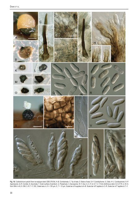
Damm et al. Fig. 16. <strong>Colletotrichum</strong> petchii (from ex-epitype strain <strong>CBS</strong> 378.94). A–B. Conidiomata. C. Tip of seta. D. Basis of seta. E–F. Conidioph<strong>or</strong>es. G. Seta. H–J. Conidioph<strong>or</strong>es. K–P. Appress<strong>or</strong>ia. Q–R. Conidia. S. Ascomata. T. Outer surface of peridium. U. Paraphysis. V. Ascosp<strong>or</strong>es. W–Y. Asci. A, C–F, Q, S–T, V, Y. from Anthriscus stem. B, G–P, R, U, W–X. from SNA. A–B, S. DM, C–R, T–Y. DIC, Scale bars: A, S = 100 µm, E, T = 10 µm. Scale bar of A applies to A–B. Scale bar of E applies to C–R. Scale bar of T applies to T–Y. 30
The <strong>Colletotrichum</strong> boninense <strong>species</strong> <strong>complex</strong> also larger than those of C. karstii and C. phyllanthi. Conidia of C. hippeastri are larger, while C. dracaenophilum occurs on Dracaena spp. as well, but is not closely related to C. petchii as demonstrated by Farr et al. (2006). Their study included <strong>CBS</strong> 118193 and <strong>CBS</strong> 118774 (C. petchii). Another <strong>species</strong> from Dracaena, C. dracaenaefragrantis, has narrower conidia, measuring 5–12 × 2.5–3.5 µm (Saccardo 1895); its affinities are unclear. <strong>Colletotrichum</strong> dracaenicola (syn. C. dracaenae Trinchieri 1909, non Allesch.) may be a synonym of C. dracaenae. The conidial size was given as 12–19 × 2–7 µm by Saccardo & Trotter (1913), which is an unusually wide range, but which overlaps with that of C. dracaenae Allesch. Farr et al. (2006) could not locate the type specimen (it was not present in NAP, PORUN <strong>or</strong> PAD), and the name theref<strong>or</strong>e remains uncertain. Von Arx (1957) considered both C. dracaenae Allesch. and C. dracaenae Petch to be synonyms of C. gloeosp<strong>or</strong>ioides. Farr et al. (2006) agreed with this conclusion concerning C. dracaenae Allesch. after studying type material, although their focus was on the need to demonstrate distinctions between C. dracaenae and their new <strong>species</strong> C. dracaenophilum. The shape of the conidia of C. dracaenae is similar to C. gloeosp<strong>or</strong>ioides, including the overall length and the constriction in the central part. The conidia were found to be noticeably wider in C. dracaenae compared with “typical” C. gloeosp<strong>or</strong>ioides. The <strong>or</strong>iginal description (Allescher 1902) of the conidia of C. dracaenae Allesch. (14–18 × 5–7 µm, elongate-cylindrical, both sides round) fits well with the <strong>species</strong> as circumscribed here. Most features of the setae also agree (40–60 µm long, obtuse tip, few septa, appearing late at the margins of conidiomata), apart from their diameter (2.5–3.5 µm acc<strong>or</strong>ding to Allescher, 5–10 µm diam as measured here in the <strong>CBS</strong> strains). In the type material, we found that the conidia measured (12.5–)13.5–16 × 5–6 µm (n = 20, mean ± SD = 15.2 ± 1.1 × 5.4 ± 0.3 µm, L/W ratio 2.8), which is smaller than those of the epitype of C. petchii but with a comparable L/W ratio. Few conidia had a noticeably prominent hilum, and the setae were found to be narrow (as observed by Allescher) and slightly verruculose. It is not certain that Allescher’s collection and the <strong>CBS</strong> isolates represent the same <strong>species</strong>, as comparisons with dried material and living cultures are difficult. As the conidial hilum m<strong>or</strong>phology seems to diverge from that seen in C. petchii (a diagnostic feature of the C. boninense aggregate) we have chosen not to use Allescher’s name. Part of the type of C. dracaenae Petch was examined by Farr et al. (2006), who noted that the fruit bodies had a very thin subhymenial layer that is only one <strong>or</strong> two layers thick. No other observations were made, and it is possible that the material they examined was effete. We re-examined the type and found conidiomata typical of the C. boninense aggregate. In conc<strong>or</strong>dance with Petch’s <strong>or</strong>iginal description, the setae are strongly curved and tapering, and strongly verruculose towards the tip. Few conidia were seen and those present were variable in shape and length/ width ratio. The maj<strong>or</strong>ity of those examined were 14–16 × 5–6.5 µm in size, and were cylindrical to doliif<strong>or</strong>m with a rather prominent hilum. We place Petch’s illegitimate taxon with confidence in the C. boninense aggregate, and it is not unreasonable to suppose that it is synonymous with C. dracaenae Allesch. Petch (1925) contrasted his <strong>species</strong> with C. c<strong>or</strong>dylines Pollacci but was evidently unaware of Allescher’s w<strong>or</strong>k. In contrast to other <strong>species</strong> in the C. boninense <strong>complex</strong>, C. dracaenae may be host-specific to Dracaena. The maj<strong>or</strong>ity of Dracaena <strong>species</strong> are native to Africa, with a few in southern Asia and one in tropical Central America, and they are often grown as pot plants <strong>or</strong> in greenhouses. The host <strong>species</strong> of the isolates studied here are popular houseplants. <strong>Colletotrichum</strong> dracaenae was mostly isolated from leaves, where it caused leaf spots as indicated in the sampling details of some of the isolates (Di Lenna & Montecchio 1995). Within the <strong>species</strong> there is only low sequence variability, and separate clusters are obtained with all phylogenies employing single genes. <strong>Colletotrichum</strong> phyllanthi (H. Surendranath Pai) Damm, P.F. Cannon & Crous, comb. nov. MycoBank MB560746. Basionym: Glomerella phyllanthi H. Surendranath Pai, Mycopath. Mycol. appl. 42: 70. 1970. Culture characteristics: Colonies on SNA flat with entire margin, hyaline, lacking aerial mycelium; reverse filter paper very pale luteous; 21.3–23.8 mm in 7 d (32.5–33.8 mm in 10 d). Colonies on OA flat with entire to slightly undulate margin; surface buff to saffron, lacking aerial mycelium, reverse same colours; 19–23 mm in 7 d (30.8–35 mm in 10 d). Anam<strong>or</strong>ph and teleom<strong>or</strong>phic structures not observed in the culture available. Material examined: India, Maharashtra, Poona, from leaf anthracnose on Phyllanthus acidus, 10 Feb. 1966, H. Surendranath Pai, IMI 122826, holotype; Maharashtra, Poona, isolated from anthracnose symptoms on leaves of Phyllanthus acidus, 10 Feb. 1966, H. Surendranath Pai, <strong>CBS</strong> H-7188, isotype, dried culture (PDA) of ascigerous stage, culture ex-isotype <strong>CBS</strong> 175.67 = MACS 271. Notes: Glomerella phyllanthi is known only from the <strong>or</strong>iginal collection taken from leaves of Phyllanthus acidus in India. The extype strain <strong>CBS</strong> 175.67, deposited in the <strong>CBS</strong> collection, did not sp<strong>or</strong>ulate under standard growth conditions. The description below is derived from the <strong>or</strong>iginal publication (Pai 1970). “Perithecia isolated <strong>or</strong> gregarious, dark brown, 159–190.8 µm with long beaks measuring 47.7–150 µm. Ostiolar threads absent. Asci numerous, unitunicate, clavate, octosp<strong>or</strong>ous, arising in basal layers, sessile to subsessile, 43.2–56.6 × 8.6–10.8 µm. Paraphyses abundant in early stages but disintegrating at maturity. Ascosp<strong>or</strong>es uniseriate <strong>or</strong> irregularly biseriate, elliptical to slightly curved, hyaline with oil globules at both ends, 12.9–17.28 × 2.1–6.4 µm.” Ascosp<strong>or</strong>e measurements from the isotype (<strong>CBS</strong> H-7188) agree with those of the <strong>or</strong>iginal description: (14–)14.5–17(–18) × (4–)4.5– 5.5(–6) µm, mean ± SD = 15.7 ± 1.1 × 5.1 ± 0.6 µm, L/W ratio = 3.1. Pai (1970) assumed that G. phyllanthi was the teleom<strong>or</strong>ph of <strong>Colletotrichum</strong> heveae, and did not provide a complete description of the anam<strong>or</strong>ph, providing the following inf<strong>or</strong>mation: acervuli 113– 159 µm, setae (only f<strong>or</strong>med in old cultures) 63–143 µm, conidia cylindrical, oblong, 14–17 × 3–5 µm. No anam<strong>or</strong>ph structures could be observed in the holotype <strong>or</strong> isotype specimens. Acc<strong>or</strong>ding to its <strong>or</strong>iginal description, conidia of Glomerella phyllanthi are narrower than the other <strong>species</strong> within the C. boninense <strong>complex</strong> and none f<strong>or</strong>med ascomata with a long beak as rep<strong>or</strong>ted from G. phyllanthi, though it must be recognised that culture medium and growth conditions were not the same. Acc<strong>or</strong>ding to the multigene phylogeny, G. phyllanthi f<strong>or</strong>ms a separate lineage close to C. karstii. This was the situation also in 5 of 7 single-gene phylogenies. Glomerella phyllanthi causes an anthracnose disease on leaves of Phyllanthus acidus in India (Pai 1966) but has not been rep<strong>or</strong>ted since. Farr & Rossman (2011) list C. gloeosp<strong>or</strong>ioides from Phyllanthus emblica in China (Zhuang 2001) and P. reticulatus in Myanmar (Thaung 2008) as well as an unidentified <strong>Colletotrichum</strong> sp. from P. acidus in India (Mathur 1979), of which at least the latter could be identical with G. phyllanthi. www.studiesinmycology.<strong>or</strong>g 31