

Pathology of the Head and Neck
Pathology of the Head and Neck
Pathology of the Head and Neck
- No tags were found...

Create successful ePaper yourself
Turn your PDF publications into a flip-book with our unique Google optimized e-Paper software.
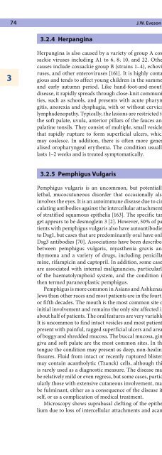
74 J.W. Eveson3.2.4 Herpangina3Herpangina is also caused by a variety <strong>of</strong> group A coxsackieviruses including A1 to 6, 8, 10, <strong>and</strong> 22. O<strong>the</strong>rcauses include coxsackie group B (strains 1–4), echoviruses,<strong>and</strong> o<strong>the</strong>r enteroviruses [161]. It is highly contagious<strong>and</strong> tends to affect young children in <strong>the</strong> summer<strong>and</strong> early autumn period. Like h<strong>and</strong>-foot-<strong>and</strong>-mouthdisease, it rapidly spreads through close-knit communities,such as schools, <strong>and</strong> presents with acute pharyngitis,anorexia <strong>and</strong> dysphagia, with or without cervicallymphadenopathy. Typically, <strong>the</strong> lesions are restricted to<strong>the</strong> s<strong>of</strong>t palate, uvula, anterior pillars <strong>of</strong> <strong>the</strong> fauces <strong>and</strong>palatine tonsils. They consist <strong>of</strong> multiple, small vesiclesthat rapidly rupture to form superficial ulcers, whichmay coalesce. In addition, <strong>the</strong>re is <strong>of</strong>ten more generalisedoropharyngeal ery<strong>the</strong>ma. The condition usuallylasts 1–2 weeks <strong>and</strong> is treated symptomatically.3.2.5 Pemphigus VulgarisFig. 3.2. Pemphigus vulgaris showing suprabasal clefting <strong>and</strong> acantholyticcells in an intraepi<strong>the</strong>lial blisterPemphigus vulgaris is an uncommon, but potentiallylethal, mucocutaneous disorder that occasionally alsoinvolves <strong>the</strong> eyes. It is an autoimmune disease due to circulatingantibodies against <strong>the</strong> intercellular attachments<strong>of</strong> stratified squamous epi<strong>the</strong>lia [163]. The specific targetappears to be desmoglein 3 [2]. However, 50% <strong>of</strong> patientswith pemphigus vulgaris also have autoantibodiesto Dsg1, but cases that are predominantly oral have onlyDsg3 antibodies [70]. Associations have been describedbetween pemphigus vulgaris, myas<strong>the</strong>nia gravis <strong>and</strong>thymoma <strong>and</strong> a variety <strong>of</strong> drugs, including penicillamine,rifampicin <strong>and</strong> captopril. In addition, some casesare associated with internal malignancies, particularly<strong>of</strong> <strong>the</strong> haematolymphoid system, <strong>and</strong> <strong>the</strong> condition is<strong>the</strong>n termed paraneoplastic pemphigus.Pemphigus is more common in Asians <strong>and</strong> AshkenaziJews than o<strong>the</strong>r races <strong>and</strong> most patients are in <strong>the</strong> fourthor fifth decades. The mouth is <strong>the</strong> most common site <strong>of</strong>initial involvement <strong>and</strong> remains <strong>the</strong> only site affected inabout half <strong>of</strong> patients. The oral features are very variable.It is uncommon to find intact vesicles <strong>and</strong> most patientspresent with painful, ragged superficial ulcers <strong>and</strong> areas<strong>of</strong> boggy <strong>and</strong> shredded mucosa. The buccal mucosa, gingiva<strong>and</strong> s<strong>of</strong>t palate are <strong>the</strong> most common sites. In <strong>the</strong>tongue <strong>the</strong> condition may present as deep, non-healingfissures. Fluid from intact or recently ruptured blistersmay contain acantholytic ( Tzanck) cells, although thisis rarely used as a diagnostic measure. The disease maybe relatively mild or even regress, but some cases, particularlythose with extensive cutaneous involvement, maybe fulminant, ei<strong>the</strong>r as a consequence <strong>of</strong> <strong>the</strong> disease itself,or as a complication <strong>of</strong> medical treatment.Microscopy shows suprabasal clefting <strong>of</strong> <strong>the</strong> epi<strong>the</strong>liumdue to loss <strong>of</strong> intercellular attachments <strong>and</strong> acantholysis(Fig 3.2). A single layer <strong>of</strong> keratinocytes may remainattached to <strong>the</strong> corium by <strong>the</strong>ir hemidesmosomes,but <strong>the</strong> cells are separated from each o<strong>the</strong>r laterally t<strong>of</strong>orm a characteristic “tombstone” appearance. The acantholyticcells floating in <strong>the</strong> vesicular fluid are rounded,with condensed cytoplasm surrounding hyperchromaticnuclei. The vesicles may contain acute <strong>and</strong> chronicinflammatory cells, <strong>and</strong> eosinophils may be a conspicuousfeature. In many cases, <strong>the</strong> ro<strong>of</strong> <strong>of</strong> <strong>the</strong> blister is lostduring <strong>the</strong> biopsy procedure due to a positive Nikolskyphenomenon, but a row <strong>of</strong> keratinocytes remains adherentto <strong>the</strong> floor. Direct immun<strong>of</strong>luorescence on frozentissue shows deposits <strong>of</strong> IgG <strong>and</strong> less frequently IgM <strong>and</strong>IgA in <strong>the</strong> intercellular junctions, producing a characteristic“chicken wire” appearance.3.2.6 Pemphigus VegetansPemphigus vegetans is considerably less common in <strong>the</strong>mouth than pemphigus vulgaris [15]. It usually presentsclinically as serpiginous ulcers that are most frequent on<strong>the</strong> dorsum <strong>of</strong> <strong>the</strong> tongue <strong>and</strong> lips [187]. The lingual lesionsclosely resemble those <strong>of</strong> ery<strong>the</strong>ma migrans. Thepapillomatous, proliferative lesions that characterisecutaneous pemphigus vegetans can sometimes be seenat <strong>the</strong> angles <strong>of</strong> <strong>the</strong> mouth. As in pemphigus vulgaris,drugs, particularly ACE inhibitors, have been invokedas possible causative agents in some cases [12, 137].Microscopically, <strong>the</strong> epi<strong>the</strong>lium tends to proliferate<strong>and</strong> become verruciform. Acantholytic cells may not beconspicuous <strong>and</strong> eosinophil microabscesses are said tobe <strong>the</strong> most typical histological feature. However, <strong>the</strong>lesions frequently resemble pyostomatitis vegetans <strong>and</strong>conventional microscopy may not be diagnostic. Thepresence <strong>of</strong> typical skin lesions <strong>of</strong>ten helps in making<strong>the</strong> diagnosis <strong>and</strong> it may be differentiated from pyostomatitisby <strong>the</strong> clinical picture <strong>and</strong> appropriate immunocytochemicalinvestigations [73].


