

Pathology of the Head and Neck
Pathology of the Head and Neck
Pathology of the Head and Neck
- No tags were found...

Create successful ePaper yourself
Turn your PDF publications into a flip-book with our unique Google optimized e-Paper software.
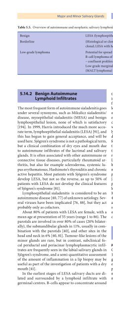
Major <strong>and</strong> Minor Salivary Gl<strong>and</strong>s Chapter 5 163Table 5.5. Overview <strong>of</strong> autoimmune <strong>and</strong> neoplastic salivary lymphoid proliferations, adapted from Quintana et al. [165]BenignBorderlineLow-grade lymphomaLESA (lymphoepi<strong>the</strong>lial sialadenitis), non-clonal(Histological or clonal evidence <strong>of</strong> neoplasia, but unlikely to disseminate): LESA,clonal; LESA with halos <strong>of</strong> marginal zone B-cellsPotential for spread to nodes <strong>and</strong> less <strong>of</strong>ten, systemically: low-grade marginal zoneB-cell lymphoma <strong>of</strong> mucosa-associated lymphoid tissue (MALT lymphoma)– confluent proliferation <strong>of</strong> marginal zone B-cellsLow-grade marginal zone B-cell lymphoma <strong>of</strong> mucosa-associated lymphoid tissue(MALT lymphoma) with plasmacytic differentiation5.14.2 Benign AutoimmuneLymphoid InfiltratesThe most frequent form <strong>of</strong> autoimmune sialadenitis goesunder several synonyms, such as Mikulicz sialadenitis/disease, myoepi<strong>the</strong>lial sialadenitis (MESA) <strong>and</strong> benignlymphoepi<strong>the</strong>lial lesion, none <strong>of</strong> which is satisfactory[194]. In 1999, Harris introduced <strong>the</strong> much more accurateterm, lymphoepi<strong>the</strong>lial sialadenitis (LESA) [91], <strong>and</strong>this has begun to gain general acceptance, <strong>and</strong> will beused here. Sjögren’s syndrome is not a pathological term,but a clinical combination <strong>of</strong> dry eyes <strong>and</strong> mouth dueto autoimmune infiltrates <strong>of</strong> <strong>the</strong> lacrimal <strong>and</strong> salivarygl<strong>and</strong>s. It is <strong>of</strong>ten associated with o<strong>the</strong>r autoimmune orconnective tissue diseases, particularly rheumatoid arthritis,but also for example scleroderma, systemic lupusery<strong>the</strong>matosus, Hashimoto’s thyroiditis <strong>and</strong> chronicactive hepatitis. Most patients with Sjögren’s syndromedevelop LESA, but not so <strong>the</strong> reverse, as up to 50% <strong>of</strong>patients with LESA do not develop <strong>the</strong> clinical features<strong>of</strong> Sjögren’s syndrome [81].Lymphoepi<strong>the</strong>lial sialadenitis is considered to be anautoimmune disease [40, 77] <strong>of</strong> unknown aetiology. Severalviruses have been implicated [76, 88], but <strong>the</strong>y actprobably only as c<strong>of</strong>actors.About 80% <strong>of</strong> patients with LESA are female, with amean age at presentation <strong>of</strong> 55 years (range 1 to 86). Theparotids are involved in over 80% <strong>of</strong> cases (20% bilaterally),<strong>the</strong> subm<strong>and</strong>ibular gl<strong>and</strong>s in 11%, usually in combinationwith <strong>the</strong> parotids [40], <strong>and</strong> o<strong>the</strong>r sites in <strong>the</strong>head <strong>and</strong> neck in 6% [40, 81]. Tumour-like lesions <strong>of</strong> <strong>the</strong>minor gl<strong>and</strong>s are rare, but in contrast, subclinical focalperiductal <strong>and</strong> periacinar lymphoplasmacytic infiltratesare frequently seen in <strong>the</strong> labial salivary gl<strong>and</strong>s inSjögren’s syndrome, <strong>and</strong> a semi-quantitative assessment<strong>of</strong> <strong>the</strong> amount <strong>of</strong> inflammation in a lip biopsy may beuseful as part <strong>of</strong> <strong>the</strong> investigation <strong>of</strong> patients with a drymouth [41].In <strong>the</strong> earliest stages <strong>of</strong> LESA salivary ducts are dilated<strong>and</strong> surrounded by a lymphoid infiltrate withgerminal centres. B-cells appear to concentrate around<strong>the</strong> ducts <strong>and</strong> focally infiltrate <strong>the</strong> epi<strong>the</strong>lium, unlikein many non-autoimmune chronic inflammatory infiltrates,where lymphocytic invasion <strong>of</strong> <strong>the</strong> ducts isless marked. In LESA many B-cells are <strong>of</strong> monocytoidor marginal zone type [91]. They are slightly largerthan small lymphocytes <strong>of</strong> <strong>the</strong> mantle zone, <strong>and</strong> arecharacterised by nuclei with irregular outlines somewhatresembling centrocytes [105]. Plasma cells <strong>and</strong> T-lymphocytes are also present. In time, <strong>the</strong> ducts condense,with partial or complete loss <strong>of</strong> <strong>the</strong>ir lumina, t<strong>of</strong>orm lymphoepi<strong>the</strong>lial lesions (LELs) [105, 172, 194];<strong>the</strong>se were previously inaccurately called epimyoepi<strong>the</strong>lialisl<strong>and</strong>s <strong>and</strong> similarly, <strong>the</strong> old usage <strong>of</strong> LEL torefer to <strong>the</strong> whole lesion should be discontinued [91].LELs consist <strong>of</strong> cohesive aggregates <strong>of</strong> epi<strong>the</strong>lium withhyalinised basal lamina material containing variablenumbers <strong>of</strong> B-cells (Fig. 5.55). Myoepi<strong>the</strong>lial cells arepresent as well, but are <strong>of</strong>ten relatively inconspicuous[42]. As <strong>the</strong> disease progresses <strong>the</strong> acini become atrophied<strong>and</strong> are <strong>the</strong>n totally replaced by lymphoid tissueleading to clinical enlargement <strong>of</strong> <strong>the</strong> salivary gl<strong>and</strong>s.Monoclonality by PCR can be demonstrated in over40% <strong>of</strong> patients with LESA [165], but this alone is probablyinsufficient for a diagnosis <strong>of</strong> lymphoma, <strong>and</strong>stronger evidence is required from <strong>the</strong> demonstration<strong>of</strong> monoclonality by immunohistochemistry or flowcytometry [107].5.14.3 MalignantLymphomaICD-O:9590/3Overall, extranodal <strong>and</strong> nodal lymphomas represented16.3% <strong>of</strong> all malignant tumours <strong>of</strong> <strong>the</strong> major salivarygl<strong>and</strong>s at <strong>the</strong> AFIP from 1985 to 1995 [62]. Almost allextranodal lymphomas are marginal zone B-cell lymphoma(MALT lymphomas), which is <strong>the</strong> preferred terminology<strong>of</strong> <strong>the</strong> current WHO classification <strong>of</strong> lymphomas[108].


