- Page 2:
Antonio Cardesa · Pieter J. Slootw
- Page 6:
Professor Dr. Antonio CardesaDepart
- Page 10:
ForewordPathology of the Head and N
- Page 14:
Contents1 Benign and Potentially Ma
- Page 18:
ContentsXIII2.11.10 Low-Grade Sinon
- Page 22:
ContentsXV5.5.5 Tissue ChangesFollo
- Page 26:
ContentsXVII7.6.4 Paraganglioma . .
- Page 30:
ContentsXIX10.4 Intraocular Tissues
- Page 34:
XXIIList of ContributorsPieter J. S
- Page 38:
2 N. Gale · N. Zidar11.1 Squamous
- Page 42:
4 N. Gale · N. Zidar1An exact and
- Page 48:
Lesions of Squamous Epithelium Chap
- Page 52:
Lesions of Squamous Epithelium Chap
- Page 56:
Lesions of Squamous Epithelium Chap
- Page 60:
Lesions of Squamous Epithelium Chap
- Page 64:
Lesions of Squamous Epithelium Chap
- Page 68:
Lesions of Squamous Epithelium Chap
- Page 72:
Lesions of Squamous Epithelium Chap
- Page 76:
Lesions of Squamous Epithelium Chap
- Page 80:
Lesions of Squamous Epithelium Chap
- Page 84:
Lesions of Squamous Epithelium Chap
- Page 88:
Lesions of Squamous Epithelium Chap
- Page 92:
Lesions of Squamous Epithelium Chap
- Page 96:
Lesions of Squamous Epithelium Chap
- Page 100:
Lesions of Squamous Epithelium Chap
- Page 104:
Lesions of Squamous Epithelium Chap
- Page 108:
Lesions of Squamous Epithelium Chap
- Page 112:
Chapter 2Nasal Cavityand Paranasal
- Page 116:
Nasal Cavity and Paranasal Sinuses
- Page 120:
Nasal Cavity and Paranasal Sinuses
- Page 124:
Nasal Cavity and Paranasal Sinuses
- Page 128:
Nasal Cavity and Paranasal Sinuses
- Page 132:
Nasal Cavity and Paranasal Sinuses
- Page 136:
Nasal Cavity and Paranasal Sinuses
- Page 140:
Nasal Cavity and Paranasal Sinuses
- Page 144:
Nasal Cavity and Paranasal Sinuses
- Page 148:
Nasal Cavity and Paranasal Sinuses
- Page 152:
Nasal Cavity and Paranasal Sinuses
- Page 156:
Nasal Cavity and Paranasal Sinuses
- Page 160:
Nasal Cavity and Paranasal Sinuses
- Page 164:
Nasal Cavity and Paranasal Sinuses
- Page 168:
Nasal Cavity and Paranasal Sinuses
- Page 172:
Nasal Cavity and Paranasal Sinuses
- Page 176:
Chapter 3Oral Cavity3J.W. EvesonCon
- Page 180:
Oral Cavity Chapter 3 73affected ar
- Page 184:
Oral Cavity Chapter 3 753.2.7 Paran
- Page 188:
Oral Cavity Chapter 3 77are present
- Page 192:
Oral Cavity Chapter 3 79Fig. 3.5. M
- Page 196:
Oral Cavity Chapter 3 81these do no
- Page 200:
Oral Cavity Chapter 3 83Fig. 3.9. L
- Page 204:
Oral Cavity Chapter 3 85keratosis o
- Page 208:
Oral Cavity Chapter 3 87Fig. 3.12.
- Page 212:
Oral Cavity Chapter 3 89tropic horm
- Page 216:
Oral Cavity Chapter 3 91Fig. 3.16.
- Page 220:
Oral Cavity Chapter 3 93erable inte
- Page 224:
Oral Cavity Chapter 3 953.7.3 Verru
- Page 228:
Oral Cavity Chapter 3 97ynx. It is
- Page 232:
Oral Cavity Chapter 3 9935. Chaudhr
- Page 236:
Oral Cavity Chapter 3 101136. Pindb
- Page 240:
Chapter 4Maxillofacial Skeletonand
- Page 244:
Maxillofacial Skeleton and Teeth Ch
- Page 248:
Maxillofacial Skeleton and Teeth Ch
- Page 252:
Maxillofacial Skeleton and Teeth Ch
- Page 256:
Maxillofacial Skeleton and Teeth Ch
- Page 260:
Maxillofacial Skeleton and Teeth Ch
- Page 264:
Maxillofacial Skeleton and Teeth Ch
- Page 268:
Maxillofacial Skeleton and Teeth Ch
- Page 272:
Maxillofacial Skeleton and Teeth Ch
- Page 276:
Maxillofacial Skeleton and Teeth Ch
- Page 280:
Maxillofacial Skeleton and Teeth Ch
- Page 284:
Maxillofacial Skeleton and Teeth Ch
- Page 288:
Maxillofacial Skeleton and Teeth Ch
- Page 292:
Maxillofacial Skeleton and Teeth Ch
- Page 296:
Chapter 5Major and MinorSalivary Gl
- Page 300:
Major and Minor Salivary Glands Cha
- Page 304:
Major and Minor Salivary Glands Cha
- Page 308:
Major and Minor Salivary Glands Cha
- Page 312:
Major and Minor Salivary Glands Cha
- Page 316:
Major and Minor Salivary Glands Cha
- Page 320:
Major and Minor Salivary Glands Cha
- Page 324:
Major and Minor Salivary Glands Cha
- Page 328:
Major and Minor Salivary Glands Cha
- Page 332:
Major and Minor Salivary Glands Cha
- Page 336: Major and Minor Salivary Glands Cha
- Page 340: Major and Minor Salivary Glands Cha
- Page 344: Major and Minor Salivary Glands Cha
- Page 348: Major and Minor Salivary Glands Cha
- Page 352: Major and Minor Salivary Glands Cha
- Page 356: Major and Minor Salivary Glands Cha
- Page 360: Major and Minor Salivary Glands Cha
- Page 364: Major and Minor Salivary Glands Cha
- Page 368: Major and Minor Salivary Glands Cha
- Page 372: Major and Minor Salivary Glands Cha
- Page 376: Chapter 6Nasopharynxand Waldeyer’
- Page 380: Nasopharynx and Waldeyer’s Ring C
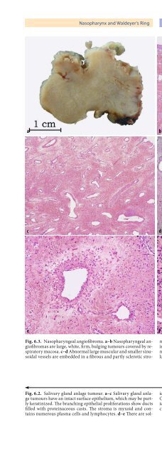
- Page 384: Nasopharynx and Waldeyer’s Ring C
- Page 390: 178 S. Regauer6sphenopalatine foram
- Page 394: 180 S. Regauer6lated and occluded e
- Page 398: 182 S. Regauer6EBV in paraffin sect
- Page 402: 184 S. Regauer6ery and within the s
- Page 406: 186 S. Regauer6abcdeFig. 6.6. EBV t
- Page 410: 188 S. Regauer6abcdeFig. 6.7. Lymph
- Page 414: 190 S. Regauer66.3.6.2 Extranodal M
- Page 418: 192 S. Regauer624. Chan JK, Yip TT,
- Page 422: 194 S. Regauer6109. Kieff DA, Curti
- Page 426: 196 S. Regauer200. Winther B, Innes
- Page 430: 198 N. Gale · A. Cardesa · N. Zid
- Page 434: 200 N. Gale · A. Cardesa · N. Zid
- Page 438:
202 N. Gale · A. Cardesa · N. Zid
- Page 442:
204 N. Gale · A. Cardesa · N. Zid
- Page 446:
206 N. Gale · A. Cardesa · N. Zid
- Page 450:
208 N. Gale · A. Cardesa · N. Zid
- Page 454:
210 N. Gale · A. Cardesa · N. Zid
- Page 458:
212 N. Gale · A. Cardesa · N. Zid
- Page 462:
214 N. Gale · A. Cardesa · N. Zid
- Page 466:
216 N. Gale · A. Cardesa · N. Zid
- Page 470:
218 N. Gale · A. Cardesa · N. Zid
- Page 474:
220 N. Gale · A. Cardesa · N. Zid
- Page 478:
222 N. Gale · A. Cardesa · N. Zid
- Page 482:
224 N. Gale · A. Cardesa · N. Zid
- Page 486:
226 N. Gale · A. Cardesa · N. Zid
- Page 490:
228 N. Gale · A. Cardesa · N. Zid
- Page 494:
230 N. Gale · A. Cardesa · N. Zid
- Page 498:
232 N. Gale · A. Cardesa · N. Zid
- Page 502:
234 N. Gale · A. Cardesa · N. Zid
- Page 506:
236 L. Michaels88.1 Summary of Embr
- Page 510:
238 L. Michaels8.2.1.4 Relapsing Po
- Page 514:
240 L. Michaelsof the oculoauriculo
- Page 518:
242 L. Michaels8.2.5 Malignant Neop
- Page 522:
244 L. Michaels88.3 Middle Earand M
- Page 526:
246 L. Michaels8Fig. 8.10. Acquired
- Page 530:
248 L. Michaels8tinct and predomina
- Page 534:
250 L. MichaelsTable 8.1. Schneider
- Page 538:
252 L. Michaels8Gross appearances a
- Page 542:
254 L. Michaelsdemarcation between
- Page 546:
256 L. Michaels8enter the labyrinth
- Page 550:
258 L. Michaels8Fig. 8.24. Lipoma o
- Page 558:
262 L. Michaels879. Nager GT (1975)
- Page 562:
264 M. A Luna · K. Pineda-Daboin9.
- Page 566:
266 M. A Luna · K. Pineda-Daboin9F
- Page 570:
268 M. A Luna · K. Pineda-Daboin9p
- Page 574:
270 M. A Luna · K. Pineda-DaboinFi
- Page 578:
272 M. A Luna · K. Pineda-Daboin9.
- Page 582:
274 M. A Luna · K. Pineda-Daboin9F
- Page 586:
276 M. A Luna · K. Pineda-DaboinTa
- Page 590:
278 M. A Luna · K. Pineda-Daboin9M
- Page 594:
280 M. A Luna · K. Pineda-Daboin9a
- Page 598:
282 M. A Luna · K. Pineda-Daboin98
- Page 602:
284 M.R. Canninga-Van Dijk1010.1 Su
- Page 606:
286 M.R. Canninga-Van Dijkepitheliu
- Page 610:
288 M.R. Canninga-Van Dijk10the cau
- Page 614:
290 M.R. Canninga-Van DijkFig. 10.9
- Page 618:
292 M.R. Canninga-Van DijkFig. 10.1
- Page 622:
294 M.R. Canninga-Van Dijk10Desceme
- Page 626:
296 M.R. Canninga-Van Dijk10uveal t
- Page 630:
298 M.R. Canninga-Van Dijk1010.4.4.
- Page 634:
300 M.R. Canninga-Van DijkFig. 10.2
- Page 638:
302 M.R. Canninga-Van Dijk10tomas h
- Page 642:
304 M.R. Canninga-Van DijkFig. 10.2
- Page 646:
306 M.R. Canninga-Van Dijkand fat i
- Page 650:
308 M.R. Canninga-Van Dijk1017. Che
- Page 654:
310 M.R. Canninga-Van Dijk10108. Sk
- Page 658:
312 Subject Indexcataract 297cellul
- Page 662:
314 Subject Indexof uveal tract 299
- Page 666:
316 Subject Indexsquamous odontogen