

Pathology of the Head and Neck
Pathology of the Head and Neck
Pathology of the Head and Neck
- No tags were found...

Create successful ePaper yourself
Turn your PDF publications into a flip-book with our unique Google optimized e-Paper software.
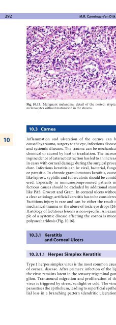
292 M.R. Canninga-Van DijkFig. 10.15. Malignant melanoma: detail <strong>of</strong> <strong>the</strong> nested, atypicalmelanocytes without maturation in <strong>the</strong> stromaFig. 10.16. Mucopolysaccharidosis: this cornea showed no abnormalitieson H&E staining, but colloidal iron showed deposits <strong>of</strong>mucopolysaccharides very clearly1010.3 CorneaInflammation <strong>and</strong> ulceration <strong>of</strong> <strong>the</strong> cornea can becaused by trauma, surgery to <strong>the</strong> eye, infectious diseases<strong>and</strong> systemic diseases. The trauma can be mechanical,chemical or caused by heat or irradiation. The increasingincidence <strong>of</strong> cataract extraction has led to an increasein cases with corneal damage during <strong>the</strong> surgical procedure.Infectious keratitis can be viral, bacterial, fungalor parasitic. In chronic granulomatous keratitis, causeslike leprosy, syphilis <strong>and</strong> tuberculosis should be considered.Especially in immunocompromised patients infectiouscauses should be excluded by additional stainslike PAS, Grocott <strong>and</strong> Gram. In corneal ulcers withouta clear aetiology, artificial keratitis has to be considered.Factitious injury is rare <strong>and</strong> can be ei<strong>the</strong>r <strong>the</strong> result <strong>of</strong>mechanical trauma or <strong>the</strong> abuse <strong>of</strong> toxic eye drops [26].Histology <strong>of</strong> factitious lesions is non-specific. An example<strong>of</strong> a systemic disease affecting <strong>the</strong> cornea is mucopolysaccharidosis(Fig. 10.16).10.3.1 Keratitis<strong>and</strong> Corneal Ulcersor punctate spots (punctate keratopathy). If <strong>the</strong> diseaserecurs, <strong>the</strong> virus may infect stromal keratocytes, causingchronic destruction with ulceration. Histologically,an early herpetic ulcer shows multinucleated epi<strong>the</strong>lialcells with intranuclear viral inclusions. DNA in situhybridisation, immunohistochemistry <strong>and</strong> PCR aremodern techniques replacing <strong>the</strong> transmission electronmicroscopy that was used to demonstrate <strong>the</strong>herpes virus particles in earlier days [49]. In end-stagedisease, epi<strong>the</strong>lial changes are no longer present; <strong>the</strong>reis just fibrosis with scarring. Often, Bowman’s layer isfocally replaced by fibrosis.10.3.1.2 Corneal UlcerationDue to Systemic DiseaseSystemic vasculitides like systemic lupus ery<strong>the</strong>matosus,polyarteritis nodosa <strong>and</strong> Wegener’s granulomatosiscan cause peripheral corneal ulceration due to vascularocclusion by immune complex deposition in <strong>the</strong>limbal vessels [73, 85]. In rheumatoid arthritis cornealulceration can occur due to <strong>the</strong> release <strong>of</strong> collagenases[92].10.3.1.1 Herpes Simplex KeratitisType 1 herpes simplex virus is <strong>the</strong> most common cause<strong>of</strong> corneal disease. After primary infection <strong>of</strong> <strong>the</strong> lip,<strong>the</strong> virus remains latent in <strong>the</strong> sensory trigeminal ganglion.Transneural migration <strong>and</strong> proliferation <strong>of</strong> <strong>the</strong>virus is triggered by stress, sunlight or cold. The virusparasitises <strong>the</strong> epi<strong>the</strong>lium, leading to superficial epi<strong>the</strong>lialloss in a branching pattern (dendritic ulceration)10.3.2 KeratoconusThis condition presents at puberty <strong>and</strong> has been foundin association with systemic disorders like Marfan’s syndrome,Down’s syndrome [52, 88], neur<strong>of</strong>ibromatosis,Ehlers-Danlos syndrome [56, 94, 129] <strong>and</strong> atopic dermatitis[93, 109, 119]. It can also be seen in combinationwith ocular disorders like aniridia, cataract <strong>and</strong> retinitispigmentosa [11, 33, 53]. A progressive, non-inflamma-


