

Pathology of the Head and Neck
Pathology of the Head and Neck
Pathology of the Head and Neck
- No tags were found...

Create successful ePaper yourself
Turn your PDF publications into a flip-book with our unique Google optimized e-Paper software.
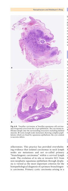
Nasopharynx <strong>and</strong> Waldeyer’s Ring Chapter 6 189lymph node or carcinoma arising in a branchiogeniccyst is probably a hypo<strong>the</strong>tical entity. Reports <strong>of</strong> a supposedbranchiogenic carcinoma included an extremelywell-differentiated SCC arising in <strong>the</strong> background<strong>of</strong> longst<strong>and</strong>ing chronic inflammation <strong>and</strong> scarring,one carcinoma arising from pre-auricular ectodermalremnants <strong>of</strong> <strong>the</strong> first pharyngeal/branchial cleft <strong>and</strong>ano<strong>the</strong>r report <strong>of</strong> a well-differentiated mucoepidermoidbranchiogenic carcinoma [16, 154, 178]. Treatment<strong>of</strong> SCC <strong>of</strong> Waldeyer’s ring is surgical resectionwith a neck dissection.6.3.6 Malignant Lymphomas<strong>of</strong> Waldeyer’s ringabFig. 6.8. Tonsillar carcinoma. a Tonsillar squamous cell carcinomasfrequently arise from <strong>the</strong> crypt epi<strong>the</strong>lium. The carcinoma infiltratesdeeply into <strong>the</strong> surrounding structures including skeletalmuscles. b Cystic lymph node metastasis showing complex papillationswhich are lined by squamous epi<strong>the</strong>lium; <strong>the</strong> cyst contentis necrotic debrissillectomies. This practice has provided overwhelmingevidence that isolated carcinomas in neck lymphnodes are metastases <strong>and</strong> not so-called primary“branchiogenic carcinomas” within a cervical lymphnode. The evolution <strong>of</strong> in situ or invasive SCC fromnon-neoplastic squamous epi<strong>the</strong>lium through dysplasiais viewed as <strong>the</strong> most important criterion for <strong>the</strong>histopathological diagnosis <strong>of</strong> a primary branchiogeniccarcinoma. Primary cystic carcinoma arising in aThis section gives a brief overview <strong>of</strong> lymphomas <strong>of</strong>Waldeyer’s ring. For a more detailed description, includinggenetic characteristics <strong>of</strong> <strong>the</strong>se lymphomassee <strong>the</strong> WHO classification <strong>and</strong> <strong>the</strong> revised European-American classification <strong>of</strong> lymphoid neoplasms [74,95]. Extranodal lymphomas <strong>of</strong> Waldeyer’s ring constituteabout 5–10% <strong>of</strong> all lymphomas in <strong>the</strong> USA <strong>and</strong>Europe, about 15% in Hong Kong <strong>and</strong> about 10–20%in Japan. Of all lymphomas involving Waldeyer’s ring,80% are primary to this site <strong>and</strong> <strong>the</strong> tonsillar fossa is<strong>the</strong> most common location, followed by <strong>the</strong> nasopharynx<strong>and</strong> <strong>the</strong> base <strong>of</strong> <strong>the</strong> tongue. Up to 20% <strong>of</strong> patientswith tonsillar lymphoma have an associated gastrointestinalinvolvement. Clinical presentation is that <strong>of</strong>a localised neoplasm, sore throat, dysphagias, <strong>and</strong> incases <strong>of</strong> nasopharyngeal involvement cranial nerve,auditory <strong>and</strong> nasal symptoms. Between 85 <strong>and</strong> 90% <strong>of</strong>all non-Hodgkin’s lymphomas in Waldeyer’s ring are<strong>of</strong> <strong>the</strong> B-cell phenotype, <strong>the</strong> remainder are <strong>of</strong> <strong>the</strong> T-celltype, but regional differences have been reported [146,150]. The vast majority <strong>of</strong> Waldeyer’s ring lymphomasare high-grade lymphomas, with only less than 15%being <strong>of</strong> low grade [113]. The majority <strong>of</strong> AIDS-relatedextranodal head <strong>and</strong> neck lymphomas are aggressiveB-cell lymphomas <strong>of</strong> <strong>the</strong> Burkitt type or immunoblasticdiffuse large B-cell lymphomas [207].6.3.6.1 Mantle Cell LymphomaICD-O:9673/3Mantle cell lymphoma (or centrocytic (mantle cell) lymphomain <strong>the</strong> Kiel classification; diffuse, small cleavedcell type in <strong>the</strong> Working Formulation) constitutes about5% <strong>of</strong> all non-Hodgkin’s B-cell lymphomas. Commonextranodal sites are <strong>the</strong> spleen, bone marrow, gastrointestinaltract <strong>and</strong> Waldeyer’s ring, particularly <strong>the</strong> palatinetonsil.


