

Pathology of the Head and Neck
Pathology of the Head and Neck
Pathology of the Head and Neck
- No tags were found...

You also want an ePaper? Increase the reach of your titles
YUMPU automatically turns print PDFs into web optimized ePapers that Google loves.
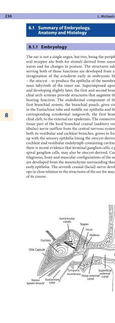
236 L. Michaels88.1 Summary <strong>of</strong> Embryology,Anatomy <strong>and</strong> Histology8.1.1 EmbryologyThe ear is not a single organ, but two, being <strong>the</strong> peripheralreceptor site both for stimuli derived from soundwaves <strong>and</strong> for changes in posture. The structures subservingboth <strong>of</strong> those functions are developed from aninvagination <strong>of</strong> <strong>the</strong> ectoderm early in embryonic life– <strong>the</strong> otocyst – to produce <strong>the</strong> epi<strong>the</strong>lia <strong>of</strong> <strong>the</strong> membranouslabyrinth <strong>of</strong> <strong>the</strong> inner ear. Superimposed upon,<strong>and</strong> developing slightly later, <strong>the</strong> first <strong>and</strong> second branchialarch systems provide structures that augment <strong>the</strong>hearing function. The endodermal component <strong>of</strong> <strong>the</strong>first branchial system, <strong>the</strong> branchial pouch, gives riseto <strong>the</strong> Eustachian tube <strong>and</strong> middle ear epi<strong>the</strong>lia <strong>and</strong> <strong>the</strong>corresponding ectodermal outgrowth, <strong>the</strong> first branchialcleft, to <strong>the</strong> external ear epidermis. The connectivetissue part <strong>of</strong> <strong>the</strong> local branchial cranial (auditory vestibular)nerve outflow from <strong>the</strong> central nervous system,both its vestibular <strong>and</strong> cochlear branches, grows to linkup with <strong>the</strong> sensory epi<strong>the</strong>lia lining <strong>the</strong> otocyst-derivedcochlear <strong>and</strong> vestibular endolymph-containing cavities;<strong>the</strong>re is recent evidence that terminal ganglion cells, e.g.spiral ganglion cells, may also be otocyst-derived. Cartilaginous,bony <strong>and</strong> muscular configurations <strong>of</strong> <strong>the</strong> earare developed from <strong>the</strong> mesenchyme surrounding <strong>the</strong>seearly epi<strong>the</strong>lia. The seventh cranial (facial) nerve developsin close relation to <strong>the</strong> structures <strong>of</strong> <strong>the</strong> ear for much<strong>of</strong> its course.8.1.2 AnatomyThe anatomy <strong>of</strong> <strong>the</strong> ear may be considered by referenceto its functions in hearing <strong>and</strong> balance (Fig. 8.1).The pinna <strong>and</strong> external canal conduct sound wavesin <strong>the</strong> air to <strong>the</strong> tympanic membrane, which transmits<strong>the</strong>m by very delicate vibrations. The middle ear enhancesthis sound energy transmission by conveying vibrationsfrom <strong>the</strong> larger area <strong>of</strong> <strong>the</strong> tympanic membranethrough <strong>the</strong> ossicular chain (malleus, incus <strong>and</strong> stapes)to <strong>the</strong> much smaller area <strong>of</strong> <strong>the</strong> footplate <strong>of</strong> <strong>the</strong> stapes,which lies in <strong>the</strong> oval window <strong>of</strong> <strong>the</strong> vestibule in contactwith <strong>the</strong> perilymph. In this way vibrations representingsound are conducted to <strong>the</strong> fluids <strong>of</strong> <strong>the</strong> inner ear. Theair space <strong>of</strong> <strong>the</strong> middle ear cavity is magnified by <strong>the</strong>mastoid air cells, which are complex expansions into <strong>the</strong>mastoid bone. There is a connection <strong>of</strong> <strong>the</strong> middle earspace with <strong>the</strong> nasopharynx <strong>and</strong> so with <strong>the</strong> external airthrough <strong>the</strong> Eustachian tube, by which air pressure canbe adjusted.From <strong>the</strong> vestibular perilymph, vibrations derivedfrom sound waves pass directly via <strong>the</strong> scala vestibuliinto <strong>the</strong> spirally coiled perilymphatic spaces <strong>of</strong> <strong>the</strong> cochlea,where it forms an upper compartment ascendingfrom <strong>the</strong> vestibule <strong>and</strong> oval window. A lower compartment,<strong>the</strong> scala tympani, descends spirally to <strong>the</strong> roundwindow membrane, a connective tissue disc separating<strong>the</strong> perilymph compartment from <strong>the</strong> middle ear. Between<strong>the</strong> scalae vestibuli <strong>and</strong> tympani <strong>the</strong>re is an endolymph-containingcoiled middle compartment, <strong>the</strong>cochlear duct (scala media), which houses <strong>the</strong> senso-Fig. 8.1. Anatomical diagram <strong>of</strong> <strong>the</strong>ear. Reproduced from Michaels <strong>and</strong>Hellquist [68]Tympanicmembrane


