Hybrid Restorations with the CAMLOG Implant System (PDF
Hybrid Restorations with the CAMLOG Implant System (PDF
Hybrid Restorations with the CAMLOG Implant System (PDF

Create successful ePaper yourself
Turn your PDF publications into a flip-book with our unique Google optimized e-Paper software.
HYBRID RESTORATIONS WITH THE <strong>CAMLOG</strong> ® IMPLANT SYSTEM<br />
PLANNING OF THE PROSTHETIC<br />
RESTORATION<br />
INTRODUCTION<br />
Modern implant pros<strong>the</strong>tics is planned by working back from <strong>the</strong> desired<br />
<strong>the</strong>rapy goal; this is referred to as "backward planning." It applies particularly<br />
to pre-implantation augmentation procedures to restore sufficient<br />
bony structure to allow placement of implants in <strong>the</strong> optimal pros<strong>the</strong>tic<br />
position.<br />
The restoration of function, phonetics and enabling good hygienic potential<br />
of <strong>the</strong> pros<strong>the</strong>sis in an edentulous arch require pros<strong>the</strong>tically oriented implant<br />
positioning and dimensioning, which <strong>the</strong> dental technician can define<br />
on <strong>the</strong> basis of <strong>the</strong> specific oral situation <strong>with</strong> a wax-up/set-up. The pros<strong>the</strong>tic<br />
design and <strong>the</strong> required implant position(s), axial alignment(s) and<br />
implant-supported anchorage options are planned and selected by <strong>the</strong> dentist<br />
and dental technician working closely toge<strong>the</strong>r. This requires both to be<br />
fully informed of <strong>the</strong> treatment options.<br />
DIAGNOSTIC CASTS, WAX-UP/SET-UP<br />
Diagnostic casts are used to represent <strong>the</strong> oral anatomical features such as<br />
<strong>the</strong> contour and size of <strong>the</strong> alveolar ridge, vestibular folds, oral bands and<br />
retromolar areas. The diagnostic casts are mounted in an adjustable articulator<br />
<strong>with</strong> <strong>the</strong> aid of an arbitrary face bow and centric registration, making<br />
it possible to represent <strong>the</strong> planned pros<strong>the</strong>tic restoration in <strong>the</strong> form of a<br />
wax set-up. The planned pros<strong>the</strong>tic result, <strong>the</strong> planned implant positions<br />
and <strong>the</strong> contour of <strong>the</strong> alveolar ridge are taken into account.<br />
DIMENSION CONTROL WITH SILICONE INDEX<br />
A silicone index created from a wax set-up is used to represent <strong>the</strong> space<br />
requirement for <strong>the</strong> planned full denture restoration on <strong>the</strong> diagnostic cast.<br />
The index should embrace <strong>the</strong> tooth arch from oral to vestibular. After curing,<br />
<strong>the</strong> index is divided along <strong>the</strong> incisal or occlusal midline. After removing<br />
<strong>the</strong> set-up, <strong>the</strong> corresponding silicone index half (buccal or palatinal/<br />
lingual half) shows <strong>the</strong> space requirement for <strong>the</strong> restoration. The silicone<br />
index can <strong>the</strong>n be used to determine optimal implant positioning, axis alignments<br />
and anchoring systems.<br />
ARCH RELATIONS<br />
The arch relations has effects on <strong>the</strong> load direction and <strong>the</strong>refore on <strong>the</strong><br />
axial alignment of <strong>the</strong> implants. This is particularly important <strong>with</strong> cross-bite<br />
situations.<br />
10<br />
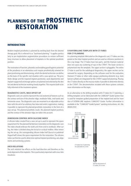
X-RAY/DRILLING TEMPLATE WITH CT-TUBES<br />
FOR CT PLANNING<br />
In a planning template fabricated on <strong>the</strong> diagnostic cast, CT-tubes are integrated<br />
at <strong>the</strong> ideal implant position and are used as reference positions in<br />
<strong>the</strong> x-ray image. The CT-tubes have two parts, and <strong>the</strong> titanium material<br />
does not cause any scattering of rays in <strong>the</strong> CT/DVT. The lower section is<br />
polymerized into <strong>the</strong> template. The upper section is pluggable. The entire<br />
CT-tube is used for <strong>the</strong> radiological diagnostics; <strong>the</strong> upper section can be<br />
removed for surgery. Depending on <strong>the</strong> software used for <strong>the</strong> evaluation,<br />
titanium CT-tubes or o<strong>the</strong>r radio-opaque positioning elements (e.g. steel,<br />
barium sulfate) are integrated for <strong>the</strong> CT/DVT-supported planning. Placing<br />
<strong>the</strong> CT-tubes directly on <strong>the</strong> mucosa makes it possible to determine density<br />
in <strong>the</strong> CT/DVT. The documentation included <strong>with</strong> <strong>the</strong>se systems contains<br />
more information on this topic.<br />
As an alternative to <strong>the</strong> drilling template <strong>with</strong> CT-tubes for CT planning, a<br />
drilling template can be fabricated <strong>with</strong> <strong>the</strong> <strong>CAMLOG</strong> ® Guide <strong>System</strong> that<br />
is used for template-guided preparation of <strong>the</strong> implant bed and for insertion<br />
of SCREW-LINE implants <strong>CAMLOG</strong> ® Guide. Fur<strong>the</strong>r information is<br />
available in <strong>the</strong> "<strong>CAMLOG</strong> ® Guide <strong>System</strong>" working instructions, Art. No.<br />
J8000.0107.<br />
10 mm<br />
Ø 2.1 mm<br />
internal diameter<br />
Ø 2.5 mm<br />
external diameter<br />
4 mm<br />
CT-tubes for CT planning<br />
for pilot drill Ø 2.0 mm<br />
Drill for placement of<br />
CT-tubes, Ø 2.0 mm