Drosophila - Severo Ochoa - Universidad Autónoma de Madrid
Drosophila - Severo Ochoa - Universidad Autónoma de Madrid
Drosophila - Severo Ochoa - Universidad Autónoma de Madrid

You also want an ePaper? Increase the reach of your titles
YUMPU automatically turns print PDFs into web optimized ePapers that Google loves.
Jefe <strong>de</strong> Línea /<br />
Group Lea<strong>de</strong>r:<br />
F. Javier Díez Guerra<br />
Bases moleculares<br />
<strong>de</strong> la plasticidad neuronal<br />
Molecular bases of neuronal<br />
plasticity<br />
D4<br />
Publicaciones<br />
Publications<br />
Resumen <strong>de</strong> investigación<br />
Research summary<br />
Rodriguez A., Durán A., Selloum M., Champy M-F, Díez-Guerra F.J.,<br />
Flores J M., Serrano M., Auwerx J., Díaz-Meco M. T. and Moscat, J.<br />
(2006). Mature-onset obesity and insulin resistance in mice <strong>de</strong>ficient<br />
in the signaling adapter p62. Cell Metabolism. 3, 211-222.<br />
Becarios Predoctorales /<br />
Predoctoral Fellows:<br />
Irene Domínguez González<br />
Alicia Algaba García<br />
Técnicos <strong>de</strong> Investigación /<br />
Technical Assistance:<br />
Silvia N. Vázquez Cuesta<br />
Neurobiología Neurobiology<br />
La plasticidad neuronal (PN) es la capacidad que tienen los<br />
contactos sinápticos <strong>de</strong> regular su eficiencia <strong>de</strong> transmisión<br />
en respuesta a la actividad que soportan. Es clave para el<br />
funcionamiento <strong>de</strong>l sistema nervioso y, muy especialmente,<br />
para la memoria, el aprendizaje y el <strong>de</strong>sarrollo y<br />
regeneración neuronales. La PN utiliza un complejo<br />
entramado <strong>de</strong> mecanismos <strong>de</strong> señalización intra y<br />
extracelulares para modificar <strong>de</strong> forma específica y local<br />
la conductancia y distribución intracelular <strong>de</strong> canales<br />
iónicos, la dinámica <strong>de</strong> citoesqueleto y morfología celular y<br />
la expresión y distribución <strong>de</strong> elementos reguladores<br />
y estructurales.<br />
Nuestro grupo se interesa en los mecanismos moleculares y<br />
celulares responsables <strong>de</strong> la PN y centra su atención en el<br />
estudio <strong>de</strong> las proteínas GMC. Esta es una familia <strong>de</strong><br />
proteínas relacionadas estructural y funcionalmente con un<br />
patrón peculiar <strong>de</strong> expresión espacio-temporal en el<br />
<strong>de</strong>sarrollo. Su interacción con proteínas <strong>de</strong> señalización<br />
intracelular y capacidad <strong>de</strong> alterar la morfología celular las<br />
asocia a PN. Sin embargo, su mecanismo <strong>de</strong> acción se<br />
<strong>de</strong>sconoce. Nuestro grupo <strong>de</strong>sarrolla y utiliza mo<strong>de</strong>los<br />
celulares para analizar la expresión, localización subcelular,<br />
modificaciones e interacciones <strong>de</strong> las proteínas GMC.<br />
Nuestro interés en la dinámica <strong>de</strong> los procesos implicados<br />
nos lleva a utilizar fusiones con proteínas fluorescentes para<br />
estudiar “in vivo” la localización subcelular y analizar la<br />
relevancia <strong>de</strong> dominios y sitios <strong>de</strong> fosforilación o acilación<br />
mediante técnicas <strong>de</strong> microscopía avanzadas.<br />
En resumen, preten<strong>de</strong>mos aportar no sólo conocimiento<br />
básico sobre la funcionalidad <strong>de</strong> las proteínas GMC, sino<br />
también explorar su potencial como dianas terapéuticas en<br />
mo<strong>de</strong>los <strong>de</strong> regeneración neuronal y en la prevención <strong>de</strong><br />
neuro<strong>de</strong>generación asociada al <strong>de</strong>sarrollo y propagación<br />
<strong>de</strong> focos epilépticos.<br />
Neuronal plasticity (NP) refers to the ability of synaptic<br />
contacts to regulate its efficiency in response to the activity<br />
load that they support. NP is basic for normal operation of the<br />
nervous system and, particularly, for memory, learning and<br />
neuronal <strong>de</strong>velopment and regeneration. NP uses a complex<br />
network of intra- and extracellular signalling mechanisms to<br />
modify in a specific and local way the conductance and<br />
intracellular distribution of ion channels, cytoskeleton<br />
dynamics and cellular morphology, and the expression and<br />
distribution of regulatory and structural elements.<br />
Our group is interested in the molecular and cellular<br />
mechanisms responsible for NP and focuses its effort in the<br />
study of GMC proteins. This is a family of proteins related both<br />
structurally and functionally, with a peculiar spatio-temporal<br />
expression pattern during <strong>de</strong>velopment. They have been<br />
associated to NP based on their interaction with signalling<br />
proteins and capacity of altering cell morphology. However,<br />
their molecular mechanisms are not fully un<strong>de</strong>rstood. Our<br />
group <strong>de</strong>velops and uses cellular mo<strong>de</strong>ls to analyze the<br />
expression, subcellular localization, modifications and<br />
interactions of GMC proteins. Our interest in the dynamics of<br />
the processes involved, leads us to use fluorescent proteins<br />
fusions to study subcellular localization "in vivo" and analyze<br />
the relevance of domains and sites of phosphorylation or<br />
acylation using advanced microscopy techniques.<br />
In summary, we seek to contribute not only to the basic<br />
knowledge on the functionality of GMC proteins, but also to<br />
explore their potential as therapeutic targets in neuronal<br />
regeneration mo<strong>de</strong>ls and in the prevention of the<br />
neuro<strong>de</strong>generation associated with the <strong>de</strong>velopment and<br />
propagation of epileptic foci.<br />
CBM 2005/2006<br />
94<br />
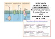
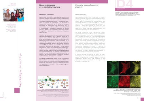
Figura 1. Esquema funcional <strong>de</strong> trabajo que ilustra la participación <strong>de</strong><br />
neurogranina en fenómenos <strong>de</strong> potenciacion / <strong>de</strong>presión (LTP / LTD) a largo plazo.<br />
Note su implicación en señalización <strong>de</strong>pendiente <strong>de</strong> calcio/calmodulina<br />
y <strong>de</strong> ácido fosfatídico.<br />
Figure 1. Functional scheme showing neurogranin (Ng) participation<br />
in long-term potentiation / <strong>de</strong>pression (LTP / LTD) in calcium / calmodulin (CaM)<br />
and phosphatidic acid (PA) <strong>de</strong>pen<strong>de</strong>nt signalling pathways.<br />
Figura 2. Células NIH-3T3 con expresión <strong>de</strong> neurogranina (rojo),<br />
fosfolipasa D2 (amarillo) y fosfatidil-inositol-4-fosfato 5-quinasa (ver<strong>de</strong>).<br />
Note el grado <strong>de</strong> colocalización entre ellas.<br />
Figure 2. NIH-3T3 cells expressing neurogranin (red), phospholipase D (yelow)<br />
and phosphatidyl-inositol-4-phosphate 5-kinase (green).<br />
Note the colocalization pattern showed.<br />
95