HIER - Congress Company
HIER - Congress Company
HIER - Congress Company

Create successful ePaper yourself
Turn your PDF publications into a flip-book with our unique Google optimized e-Paper software.
eduRAD syllabus 69<br />
28<br />
Diffusion imaging<br />
Apparent diffusion coefficient (ADC) values as measured<br />
with DWI are inversely related to cellularity, i.e. increased<br />
cellularity restricts diffusion and reduces ADC values.<br />
As increased cellularity is considered an indication of<br />
high grade tumour, it is expected that ADC may be useful<br />
for radiological tumour grading. Findings, however,<br />
are inconsistent and conflicting, with some authors<br />
reporting decrease of ADC in high grade tumours, as<br />
would be expected with increased cellularity (3), but<br />
others reporting increased ADC (4). The source of such<br />
discrepancies may, at least partially, be found in tumour<br />
heterogeneity. High grade tumours contain (micro)necrotic<br />
components in which diffusion is increased. Furthermore,<br />
diffusion is also increased in the peritumoural vasogenic<br />
oedema.<br />
MR spectroscopy<br />
Proton MR spectroscopy shows a correlation of the<br />
choline (Cho) peak with cell density and the lipid peak<br />
with proliferation (5). With increasing tumour grade there<br />
is generally an increase in the Cho/Creatine (Cr) ratio, a<br />
reduction in N-acetylaspartate (NAA) and an increase in<br />
the lactate/lipid peak (5-8). MR spectroscopy may be of<br />
particular use in the grading of oligodendrogliomas, in which<br />
contrast enhancement and increased rCBV ratios may be<br />
observed both in low and in high grades (7-9).<br />
Guiding neurosurgical intervention<br />
Among the commonly performed neurosurgical interventions<br />
we can distinguish diagnostic and therapeutic procedures.<br />
During either intervention damage to eloquent brain regions<br />
needs to be avoided. While eloquent brain regions can<br />
readily be identified on the basis of anatomical landmarks<br />
in the normal brain, such landmarks may be obscured in the<br />
presence of brain tumour with considerable mass effect.<br />
Advanced MR imaging techniques may be used to provide<br />
such information preoperatively. Furthermore, advanced MR<br />
imaging may also be used to identify the optimal target for<br />
diagnostic procedures.<br />
MR perfusion and spectroscopy<br />
Tumour grading is based on the highest malignancy grade<br />
identified within a tumour. Especially with stereotactic<br />
biopsy, sampling error is a real issue, when the tumour is<br />
under graded if the most malignant part of the tumour is<br />
not biopsied. With conventional MR imaging, stereotactic<br />
biopsy is generally targeted at the enhancing part of the<br />
tumour, which does not necessarily correspond with the<br />
most malignant part of the tumour. Such sampling errors<br />
may be avoided with the use of MR perfusion imaging or MR<br />
spectroscopy, with which the most vascular or malignant<br />
regions are readily identified.<br />
I n s c h r i j v e n v i a w w w . c o n g r e s s c o m p a n y . c o m<br />
o f w w w . r a d i o l o g e n . n l<br />
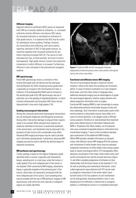
Figure 1. Combined fMRI and DTI-tractography indicating<br />
displacement of both the primary motor cortex and the corticospinal<br />
tract by the tumour mass effect.<br />
Functional and diffusion tensor MR imaging<br />
The aim of neurosurgical therapy is maximum tumour<br />
resection, while at the same time avoiding new functional<br />
deficit. In cases of tumour localisation in or near eloquent<br />
brain areas, such the motor cortex or language areas,<br />
additional advanced imaging may be advantageous to guide<br />
the neurosurgical approach, shorten surgery duration and<br />
obtain prognostic information prior to surgery.<br />
Functional MR imaging (fMRI) is used increasingly to assess<br />
the relationship between functionally eloquent cortex and<br />
brain pathology. Such information is particularly useful when<br />
normal anatomy is obscured by tumour mass effect or in<br />
cases of cortical plasticity. In an elegant study of 39 brain<br />
tumour patients, Petrella et al. demonstrated that treatment<br />
plans were altered based on information obtained with<br />
fMRI in 19 patients (10). Most notably, out of 9 patients<br />
who were considered inoperable based on information from<br />
conventional imaging, 7 were in fact considered operable<br />
upon assessment of the additional fMRI findings.<br />
While fMRI provides valuable information on eloquent<br />
cortex, with diffusion tensor MR imaging (DTI) the anatomy<br />
and involvement of white matter tracts may be evaluated.<br />
Inadvertent transection of white matter tracts during surgery<br />
leads to severe neurological deficit. DTI-tractography offers<br />
attractive visualisation of the major white matter tracts such<br />
as the corticospinal tract and the arcuate fasciculus (figure<br />
1), and offers valuable preoperative information on their<br />
relationships with the brain tumour to be resected (11-13).<br />
As well as providing such anatomical information, colour<br />
coded eigenvector maps obtained with DTI can be used<br />
to categorise involvement of the white matter tracts<br />
by brain tumour (14). Four patterns of such involvement<br />
can be distinguished, indicating whether a tract is only<br />
displaced but not infiltrated, is altered due to vasogenic<br />
oedema, is infiltrated by tumour or is completely destroyed.