- Page 1 and 2:
COMPUTATIONAL INTELLIGENCE CULTURE,
- Page 4 and 5:
The MIT Encyclopedia of the Cogniti
- Page 6:
To the memory of Henry Bradford Sta
- Page 10 and 11:
Acquisition, Formal Theories of 1 A
- Page 12 and 13:
Language Impairment, Developmental
- Page 14 and 15:
Preface The MIT Encyclopedia of the
- Page 16 and 17:
Philosophy Robert A. Wilson The are
- Page 18 and 19:
Philosophy xvii tion are constant a
- Page 20 and 21:
Philosophy xix by HELMHOLTZ, Weber,
- Page 22 and 23:
Philosophy xxi behavioral effects.
- Page 24 and 25:
Philosophy xxiii Malcolm’s point
- Page 26 and 27:
Philosophy xxv chemical kinds—suc
- Page 28 and 29:
Philosophy xxvii been to create pro
- Page 30 and 31:
Philosophy xxix Decade of the Brain
- Page 32 and 33:
Philosophy xxxi also being, to some
- Page 34 and 35:
All A are B. No B are C. No A are C
- Page 36 and 37:
Philosophy xxxv minds of other anim
- Page 38:
Philosophy xxxvii Pinker, S. (1997)
- Page 41 and 42:
xl Psychology human CHESS champion
- Page 43 and 44:
xlii Psychology Helmholtz’s insig
- Page 45 and 46:
xliv Psychology the idea that “ob
- Page 47 and 48:
xlvi Psychology the tradition of Gi
- Page 49 and 50:
xlviii Psychology in LOGIC. Althoug
- Page 52 and 53:
1 Cognitive Neuroscience Neuroscien
- Page 54 and 55:
Neurosciences liii its more pragmat
- Page 56 and 57:
Neurosciences lv activity (see ELEC
- Page 58 and 59:
Neurosciences lvii immediate object
- Page 60 and 61:
Neurosciences lix ules. At the pres
- Page 62 and 63:
Neurosciences lxi the correlation b
- Page 64 and 65:
Neurosciences lxiii ple of this typ
- Page 66 and 67:
Neurosciences lxv such as those ass
- Page 68 and 69:
Neurosciences lxvii LTP is commonly
- Page 70 and 71:
Neurosciences lxix specific cogniti
- Page 72 and 73:
Neurosciences lxxi Marr, D. (1982).
- Page 74 and 75:
Computational Intelligence Michael
- Page 76 and 77:
Computational Intelligence lxxv Bay
- Page 78 and 79:
Computational Intelligence lxxvii a
- Page 80 and 81:
Computational Intelligence lxxix et
- Page 82 and 83:
Computational Intelligence lxxxi ri
- Page 84 and 85:
Computational Intelligence lxxxiii
- Page 86 and 87:
Computational Intelligence lxxxv SU
- Page 88 and 89:
Computational Intelligence lxxxvii
- Page 90 and 91:
Computational Intelligence lxxxix 1
- Page 92 and 93:
Linguistics and Language Gennaro Ch
- Page 94 and 95:
Linguistics and Language xciii (cf.
- Page 96 and 97:
Linguistics and Language xcv follow
- Page 98 and 99:
Linguistics and Language xcvii Ther
- Page 100 and 101:
Linguistics and Language xcix wheth
- Page 102 and 103:
Linguistics and Language ci THOUGHT
- Page 104 and 105:
Linguistics and Language ciii for e
- Page 106 and 107:
Linguistics and Language cv How do
- Page 108 and 109:
Linguistics and Language cvii strea
- Page 110:
Linguistics and Language cix Chung,
- Page 113 and 114:
cxii Culture, Cognition, and Evolut
- Page 115 and 116:
cxiv Culture, Cognition, and Evolut
- Page 117 and 118:
cxvi Culture, Cognition, and Evolut
- Page 119 and 120:
cxviii Culture, Cognition, and Evol
- Page 121 and 122:
cxx Culture, Cognition, and Evoluti
- Page 123 and 124:
cxxii Culture, Cognition, and Evolu
- Page 125 and 126:
cxxiv Culture, Cognition, and Evolu
- Page 127 and 128:
cxxvi Culture, Cognition, and Evolu
- Page 129 and 130:
cxxviii Culture, Cognition, and Evo
- Page 131 and 132:
cxxx Culture, Cognition, and Evolut
- Page 133 and 134:
cxxxii Culture, Cognition, and Evol
- Page 135 and 136:
2 Acquisition, Formal Theories of l
- Page 137 and 138:
4 Affordances distinctive feature o
- Page 139 and 140:
6 Agency characteristic of any part
- Page 141 and 142:
8 Aging, Memory, and the Brain meas
- Page 143 and 144:
10 AI and Education mostly just pre
- Page 145 and 146:
12 A-Life idea that a homunculus of
- Page 147 and 148:
14 Ambiguity Seeley, T. D. (1989).
- Page 149 and 150:
16 Amygdala, Primate nuclei, each w
- Page 151 and 152:
18 Analogy issue of analogical retr
- Page 153 and 154:
20 Anaphora Winston, P. H. (1982).
- Page 155 and 156:
22 Animal Cognition Papers from the
- Page 157 and 158:
24 Animal Navigation inappropriate?
- Page 159 and 160:
26 Animal Navigation, Neural Networ
- Page 161 and 162:
28 Animism Further Readings Blair,
- Page 163 and 164:
30 Anomalous Monism Inagaki, K. (19
- Page 165 and 166:
32 Archaeology Similarly, a modalit
- Page 167 and 168:
34 Articulation diverse because no
- Page 169 and 170:
36 Artifacts and Civilization still
- Page 171 and 172:
38 Artificial Life hunting, or evas
- Page 173 and 174:
40 Attention ignore the other provi
- Page 175 and 176:
42 Attention in the Animal Brain th
- Page 177 and 178:
44 Attention in the Human Brain Fig
- Page 179 and 180:
46 Attribution Theory Stuss, D. T.,
- Page 181 and 182:
48 Audition Storms, M. D. (1973). V
- Page 183 and 184:
50 Auditory Attention Hartmann, W.
- Page 185 and 186:
52 Auditory Physiology brain measur
- Page 187 and 188:
54 Auditory Physiology Figure 1. Sc
- Page 189 and 190:
56 Auditory Plasticity thus be invo
- Page 191 and 192:
58 Autism See also AUDITION; AUDITO
- Page 193 and 194:
60 Autocatalysis Asperger, H. (1944
- Page 195 and 196:
62 Automata details and definitions
- Page 197 and 198:
64 Autonomy of Psychology been an i
- Page 199 and 200:
66 Autopoiesis Owens, D. (1989). Le
- Page 201 and 202:
68 Basal Ganglia for GPe projection
- Page 203 and 204:
70 Bayesian Learning DeVito, J. L.,
- Page 205 and 206:
72 Bayesian Networks which represen
- Page 207 and 208:
74 Behavior-Based Robotics dynamics
- Page 209 and 210:
76 Behavior-Based Robotics Referenc
- Page 211 and 212:
78 Behaviorism are dealing with act
- Page 213 and 214:
80 Belief Networks Nisbett, R. E.,
- Page 215 and 216:
82 Binding by Neural Synchrony and
- Page 217 and 218:
84 Binding by Neural Synchrony Gray
- Page 219 and 220:
86 Binding Theory References Fodor,
- Page 221 and 222:
88 Blindsight Everaert, M. (1991).
- Page 223 and 224:
90 Bloomfield, Leonard Holmes, G. (
- Page 225 and 226:
92 Boltzmann Machines Boas’s thin
- Page 227 and 228:
94 Brain Mapping Dean, T. (1991). D
- Page 229 and 230:
96 Broadbent, Donald E. Figure 1. B
- Page 231 and 232:
98 Cajal, Santiago Ramón y cogniti
- Page 233 and 234:
100 Case-Based Reasoning and Analog
- Page 235 and 236:
102 Categorial Grammar into α, whe
- Page 237 and 238:
104 Categorization Joshi, A., K. Vi
- Page 239 and 240:
106 Causal Reasoning Rips, L. J. (1
- Page 241 and 242:
108 Causation Mackie, J. L. (1974).
- Page 243 and 244:
110 Cellular Automata Strawson, G.
- Page 245 and 246:
112 Cerebral Cortex (ii) its arrang
- Page 247 and 248:
114 Chess, Psychology of have the s
- Page 249 and 250:
116 Chunking does the whole room. T
- Page 251 and 252:
118 Civilization References Church,
- Page 253 and 254:
120 Cognition and Aging Chomsky, N.
- Page 255 and 256:
122 Cognitive Archaeology Fillmore,
- Page 257 and 258:
124 Cognitive Architecture Gowlett,
- Page 259 and 260:
126 Cognitive Artifacts References
- Page 261 and 262:
128 Cognitive Development Group, Ed
- Page 263 and 264:
130 Cognitive Dissonance Further Re
- Page 265 and 266:
132 Cognitive Ethology Hollnagel, E
- Page 267 and 268:
134 Cognitive Linguistics Boden, M.
- Page 269 and 270:
136 Cognitive Maps oneself in a dif
- Page 271 and 272:
138 Cognitive Modeling, Connectioni
- Page 273 and 274:
140 Cognitive Modeling, Connectioni
- Page 275 and 276:
142 Cognitive Modeling, Symbolic th
- Page 277 and 278:
144 Color Categorization (Kay and K
- Page 279 and 280:
146 Color, Neurophysiology of reaso
- Page 281 and 282:
148 Columns and Modules (about 2% o
- Page 283 and 284:
150 Communication or repeated patch
- Page 285 and 286:
152 Competence/Performance Distinct
- Page 287 and 288:
154 Computation for example, one en
- Page 289 and 290:
156 Computation and the Brain Figur
- Page 291 and 292:
158 Computational Complexity Koch,
- Page 293 and 294:
160 Computational Lexicons will per
- Page 295 and 296:
162 Computational Linguistics appli
- Page 297 and 298:
164 Computational Neuroanatomy (pre
- Page 299 and 300:
166 Computational Neuroscience Schw
- Page 301 and 302:
168 Computational Psycholinguistics
- Page 303 and 304:
170 Computational Theory of Mind Lu
- Page 305 and 306:
172 Computational Vision Pylyshyn,
- Page 307 and 308:
174 Computer-Human Interaction Broo
- Page 309 and 310:
176 Concepts in the dendritic cable
- Page 311 and 312:
178 Concepts Five broad classes of
- Page 313 and 314:
180 Conceptual Change concept, perh
- Page 315 and 316:
182 Conceptual Role Semantics Furth
- Page 317 and 318:
184 Conditioning and the Brain Skin
- Page 319 and 320:
186 Connectionism, Philosophical Is
- Page 321 and 322:
188 Connectionist Approaches to Lan
- Page 323 and 324:
190 Consciousness items from memory
- Page 325 and 326:
192 Consciousness voluntary action,
- Page 327 and 328:
194 Consciousness, Neurobiology of
- Page 329 and 330:
196 Constraint Satisfaction exponen
- Page 331 and 332:
198 Context and Point of View Conte
- Page 333 and 334:
200 Control Theory There are import
- Page 335 and 336:
202 Cortex punishing those who chea
- Page 337 and 338:
204 Cortical Localization, History
- Page 339 and 340:
206 Creoles The Creative Person Thr
- Page 341 and 342:
208 Cross-Cultural Variation Refere
- Page 343 and 344:
210 Cultural Evolution through soci
- Page 345 and 346:
212 Cultural Psychology (Goddard 19
- Page 347 and 348:
214 Cultural Relativism such divers
- Page 349 and 350:
216 Cultural Symbolism Cultural Sym
- Page 351 and 352:
218 Culture labor: different patter
- Page 353 and 354:
220 Data Mining References Darwin,
- Page 355 and 356:
222 Decision Making The study of de
- Page 357 and 358:
224 Decision Trees known as preprun
- Page 359 and 360:
226 Deficits odological issues (see
- Page 361 and 362:
228 Depth Perception Figure 2.
- Page 363 and 364:
230 Detectors behavior of nonhuman
- Page 365 and 366:
232 Discourse behave in accordance
- Page 367 and 368:
234 Distinctive Features ory (Bem 1
- Page 369 and 370:
236 Distributed AI Gnanadesikan, A.
- Page 371 and 372:
238 Domain Specificity Földiák, P
- Page 373 and 374:
240 Dominance in Animal Social Grou
- Page 375 and 376:
242 Dreaming de Waal, F. (1989). Do
- Page 377 and 378:
244 Dynamic Approaches to Cognition
- Page 379 and 380:
246 Dynamic Programming that a dyna
- Page 381 and 382:
248 Dynamic Semantics A simple but
- Page 383 and 384:
250 Dyslexia The most common form o
- Page 385 and 386:
252 Ebbinghaus, Hermann The English
- Page 387 and 388:
254 Echolocation of fluttering inse
- Page 389 and 390:
256 Ecological Psychology the contr
- Page 391 and 392:
258 Ecological Validity determine,
- Page 393 and 394:
260 Economics and Cognitive Science
- Page 395 and 396:
262 Electric Fields computer-based
- Page 397 and 398:
264 Electrophysiology, Electric and
- Page 399 and 400:
266 Eliminative Materialism and tha
- Page 401 and 402:
268 Emergentism much less unity in
- Page 403 and 404:
270 Emotion and the Animal Brain (i
- Page 405 and 406:
272 Emotion and the Human Brain exp
- Page 407 and 408:
274 Emotions background of emotions
- Page 409 and 410:
276 Epiphenomenalism One critical r
- Page 411 and 412:
278 Episodic vs. Semantic Memory Ja
- Page 413 and 414:
280 Epistemology and Cognition Pern
- Page 415 and 416:
282 Essentialism FOLK PSYCHOLOGY, o
- Page 417 and 418:
284 Ethics Domain Specificity in Co
- Page 419 and 420:
286 Ethnopsychology beliefs; this s
- Page 421 and 422:
288 Ethology Quinn, N., and D. Holl
- Page 423 and 424:
290 Evoked Fields Craig, W. (1918).
- Page 425 and 426:
292 Evolution and Ethics themselves
- Page 427 and 428:
294 Evolutionary Computation and me
- Page 429 and 430:
296 Evolutionary Psychology about h
- Page 431 and 432:
298 Expert Systems Hirschfeld, L.,
- Page 433 and 434:
300 Explanation Djakow, I. N., N. W
- Page 435 and 436:
302 Explanation-Based Learning PROC
- Page 437 and 438:
304 Explanatory Gap Explanatory Gap
- Page 439 and 440:
306 Externalism of any component wi
- Page 441 and 442:
308 Eye Movements and Visual Attent
- Page 443 and 444:
310 Face Recognition respond to vis
- Page 445 and 446:
312 Feature Detectors ally selectiv
- Page 447 and 448:
314 Features Suga, N. (1994). Multi
- Page 449 and 450:
316 Focus for dinner, but JOHN is a
- Page 451 and 452:
318 Folk Biology (poodle, white oak
- Page 453 and 454:
320 Form/Content (von Eckardt 1994,
- Page 455 and 456:
322 Formal Systems, Properties of t
- Page 457 and 458:
324 Formal Theories Gödel, K. (193
- Page 459 and 460:
326 Frame Problem Schmidt-Schauss,
- Page 461 and 462:
328 Freud, Sigmund Dummett, M. (197
- Page 463 and 464:
330 Functional Decomposition tune,
- Page 465 and 466:
332 Functionalism when and where th
- Page 467 and 468:
334 Functionalism brains. However,
- Page 469 and 470:
336 Game-Playing Systems similarity
- Page 471 and 472:
338 Game Theory is no clear way at
- Page 473 and 474:
340 Gender equilibrium. Refinements
- Page 475 and 476:
342 Generative Grammar The second s
- Page 477 and 478:
344 Gestalt Perception (Geschwind a
- Page 479 and 480:
346 Gestalt Psychology was achieved
- Page 481 and 482:
348 Gestalt Psychology Figure 2. A
- Page 483 and 484:
350 Gibson, James Jerome observing
- Page 485 and 486:
352 Golgi, Camillo sentence; σ exp
- Page 487 and 488:
354 Good Old Fashioned AI (GOFAI) S
- Page 489 and 490:
356 Grammatical Relations object (i
- Page 491 and 492:
358 Greedy Local Search solution is
- Page 493 and 494:
360 Gustation use of or, not the wo
- Page 495 and 496:
362 Head-Driven Phrase Structure Gr
- Page 497 and 498:
364 Head Movement because they are
- Page 499 and 500:
366 Hearing See also BINDING THEORY
- Page 501 and 502:
368 Helmholtz, Hermann Ludwig Ferdi
- Page 503 and 504:
370 Hemispheric Specialization grou
- Page 505 and 506:
372 Heuristic Search Puce, A., T. A
- Page 507 and 508:
374 High-Level Vision Figure 1. A g
- Page 509 and 510:
376 High-Level Vision the world ser
- Page 511 and 512:
378 Historical Linguistics accounts
- Page 513 and 514:
380 Human Nature cognition to highl
- Page 515 and 516:
382 Human Universals advantage of s
- Page 517 and 518:
384 Hume, David Jankowiak, W. (1995
- Page 519 and 520:
386 Illusions Figure 2. Top: Illusi
- Page 521 and 522:
388 Imagery Shepard (1984) suggests
- Page 523 and 524:
390 Imitation made by another (Rizz
- Page 525 and 526:
392 Implicature Grice observes that
- Page 527 and 528:
394 Implicit vs. Explicit Memory Sa
- Page 529 and 530:
396 Indexicals and Demonstratives p
- Page 531 and 532:
398 Individualism beyond-the-head e
- Page 533 and 534:
400 Inductive Logic Programming of
- Page 535 and 536:
402 Infant Cognition structural ale
- Page 537 and 538:
404 Inference learning systems whos
- Page 539 and 540:
406 Informational Semantics symmetr
- Page 541 and 542:
408 Inheritance Fodor, J. (1984). S
- Page 543 and 544:
410 Intelligence A change in the fi
- Page 545 and 546:
412 Intentional Stance appropriate
- Page 547 and 548:
414 Intentionality my desire for wo
- Page 549 and 550:
416 Intersubjectivity (especially o
- Page 551 and 552:
418 Intersubjectivity Ecological an
- Page 553 and 554:
420 Introspection significant perio
- Page 555 and 556:
422 James, William individual langu
- Page 557 and 558:
424 Judgment Heuristics small sampl
- Page 559 and 560:
426 Justification fied beliefs are
- Page 561 and 562:
428 Kinds 4. Faculties: Kant develo
- Page 563 and 564:
430 Knowledge-Based Systems Clancey
- Page 565 and 566:
432 Knowledge Compilation MYCIN and
- Page 567 and 568:
434 Language Acquisition References
- Page 569 and 570:
436 Language Acquisition regulariza
- Page 571 and 572:
438 Language and Cognition Bloom, P
- Page 573 and 574:
440 Language and Communication pros
- Page 575 and 576:
442 Language and Gender social inte
- Page 577 and 578:
444 Language and Modularity Coates,
- Page 579 and 580:
446 Language Change Leonard, L. (19
- Page 581 and 582:
448 Language, Innateness of Curtiss
- Page 583 and 584:
450 Language, Neural Basis of Broca
- Page 585 and 586:
452 Language of Thought defenders t
- Page 587 and 588:
454 Language Production disfluencie
- Page 589 and 590:
456 Language Universals Communicati
- Page 591 and 592:
458 Lashley, Karl Spencer (1890-195
- Page 593 and 594:
460 Laws References Beach, F. A. (1
- Page 595 and 596:
462 Learning Systems matters are co
- Page 597 and 598:
464 Lexical Access two types of soc
- Page 599 and 600:
466 Lexical Functional Grammar Furt
- Page 601 and 602:
468 Lexicon type is allowed in each
- Page 603 and 604:
470 Lexicon, Neural Basis of patien
- Page 605 and 606:
472 Limbic System Figure 2. Lightne
- Page 607 and 608:
474 Limbic System basolateral limbi
- Page 609 and 610:
476 Linguistic Stress reference to
- Page 611 and 612:
478 Linguistics, Philosophical Issu
- Page 613 and 614:
480 Linguistics, Philosophical Issu
- Page 615 and 616:
482 Local Representation assumed th
- Page 617 and 618:
484 Logic Programming implicit in T
- Page 619 and 620:
486 Logical Form in Linguistics App
- Page 621 and 622:
488 Logical Form, Origins of Hornst
- Page 623 and 624:
490 Logical Omniscience, Problem of
- Page 625 and 626:
492 Long-Term Potentiation can deri
- Page 627 and 628:
494 LOT Otto, T., H. Eichenbaum, S.
- Page 629 and 630:
496 Machiavellian Intelligence Hypo
- Page 631 and 632:
498 Machine Translation models (Has
- Page 633 and 634:
500 Machine Translation documents,
- Page 635 and 636:
502 Machine Vision infer from image
- Page 637 and 638:
504 Magic and Superstition emotion
- Page 639 and 640:
506 Magnetic Resonance Imaging sinc
- Page 641 and 642:
508 Manipulation and Grasping Firth
- Page 643 and 644:
510 Manipulation and Grasping See a
- Page 645 and 646:
512 Materialism understanding brain
- Page 647 and 648:
514 Memory 4. What is the relations
- Page 649 and 650:
516 Memory This problem formed the
- Page 651 and 652:
518 Memory, Animal Studies the hipp
- Page 653 and 654:
520 Memory, Human Neuropsychology M
- Page 655 and 656:
522 Memory Storage, Modulation of i
- Page 657 and 658:
524 Mental Causation McGaugh, J. L.
- Page 659 and 660:
526 Mental Models process of compre
- Page 661 and 662:
528 Mental Representation distingui
- Page 663 and 664:
530 Mental Retardation accompanied
- Page 665 and 666:
532 Mental Rotation Figure 1. Illus
- Page 667 and 668:
534 Metacognition is usually superi
- Page 669 and 670:
536 Metaphor disaster. Sometimes a
- Page 671 and 672:
538 Metaphor and Culture the extens
- Page 673 and 674:
540 Metareasoning Figure 1. The con
- Page 675 and 676:
542 Metarepresentation has been des
- Page 677 and 678:
544 Meter and Poetry This causes a
- Page 679 and 680:
546 Mind-Body Problem Figure 3. Fig
- Page 681 and 682:
548 Mind Design How can the complex
- Page 683 and 684:
550 Minimum Description Length Econ
- Page 685 and 686:
552 Mobile Robots mining, waste rem
- Page 687 and 688:
554 Modal Logic Musliner D., E. Dur
- Page 689 and 690:
556 Modeling Neuropsychological Def
- Page 691 and 692:
558 Modularity of Mind that perhaps
- Page 693 and 694:
560 Modularity of Mind modularity m
- Page 695 and 696:
562 Morphology formal and universal
- Page 697 and 698:
564 Motion, Perception of aspects o
- Page 699 and 700:
566 Motivation Motion and Its Use i
- Page 701 and 702:
568 Motivation and Culture abstract
- Page 703 and 704:
570 Motor Control Paul, R. (1990).
- Page 705 and 706:
572 Motor Learning which muscular f
- Page 707 and 708:
574 Multisensory Integration Clearw
- Page 709 and 710:
576 Naive Mathematics people will l
- Page 711 and 712:
578 Naive Physics will follow a str
- Page 713 and 714:
580 Naive Sociology particularly in
- Page 715 and 716:
582 Narrow Content 1980; see also F
- Page 717 and 718:
584 Nativism innate, to particular
- Page 719 and 720:
586 Nativism, History of Spelke, E.
- Page 721 and 722:
588 Natural Kinds Scott, D. (1996).
- Page 723 and 724:
590 Natural Language Generation Fig
- Page 725 and 726:
592 Natural Language Processing Hov
- Page 727 and 728:
594 Navigation There are many appli
- Page 729 and 730:
596 Neural Development illustrate.
- Page 731 and 732:
598 Neural Plasticity the problem i
- Page 733 and 734:
600 Neural Plasticity cortical stag
- Page 735 and 736:
602 Neuroendocrinology produces a l
- Page 737 and 738:
604 Neuron the patterns of synaptic
- Page 739 and 740:
606 Neurotransmitters The second ne
- Page 741 and 742:
608 Nonmonotonic Logics In addition
- Page 743 and 744:
610 Nonmonotonic Logics a nonclassi
- Page 745 and 746:
612 Numeracy and Culture school (Br
- Page 747 and 748:
614 Object Recognition, Animal Stud
- Page 749 and 750:
616 Object Recognition, Human Neuro
- Page 751 and 752:
618 Oculomotor Control Whether this
- Page 753 and 754:
620 Olfaction Schiller, P. H., S. D
- Page 755 and 756:
622 Origins of Intelligence RELATIO
- Page 757 and 758:
624 Parallelism in AI of the face.
- Page 759 and 760:
626 Parameter-Setting Approaches to
- Page 761 and 762:
628 Parsimony and Simplicity Normal
- Page 763 and 764:
630 Pattern Recognition and Feedfor
- Page 765 and 766:
632 Perception neuropsychological s
- Page 767 and 768:
634 Perceptual Development (Bahrick
- Page 769 and 770:
636 Philosophical Issues in Linguis
- Page 771 and 772:
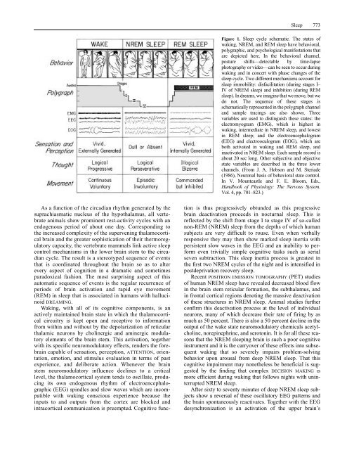
638 Phonological Rules and Processe
- Page 773 and 774:
640 Phonology tone distinguishes si
- Page 775 and 776:
642 Phonology, Acquisition of that,
- Page 777 and 778:
644 Phonology, Neural Basis of feat
- Page 779 and 780:
646 Physicalism these emergent prop
- Page 781 and 782:
648 Pictorial Art and Vision novel
- Page 783 and 784:
650 Pictorial Art and Vision crease
- Page 785 and 786:
652 Planning References McCulloch,
- Page 787 and 788:
654 Plasticity References Allen, J.
- Page 789 and 790:
656 Population-Level Cognitive Phen
- Page 791 and 792:
658 Positron Emission Tomography Fi
- Page 793 and 794:
660 Poverty of the Stimulus Argumen
- Page 795 and 796:
662 Pragmatics or token-reflexives
- Page 797 and 798:
664 Presupposition Discourse Descri
- Page 799 and 800:
666 Primate Amygdala The consequent
- Page 801 and 802:
668 Primate Cognition References Bo
- Page 803 and 804:
670 Primate Language Both Kanzi and
- Page 805 and 806:
672 Probabilistic Reasoning As a re
- Page 807 and 808:
674 Problem Solving of the new rock
- Page 809 and 810:
676 Procedural Semantics knowledge-
- Page 811 and 812:
678 Propositional Attitudes Newell,
- Page 813 and 814:
680 Prosody and Intonation Phonolog
- Page 815 and 816:
682 Prosody and Intonation, Process
- Page 817 and 818:
684 Psychoanalysis, Contemporary Vi
- Page 819 and 820:
686 Psychoanalysis, History of and
- Page 821 and 822:
688 Psycholinguistics Glymour, C. (
- Page 823 and 824:
690 Psychological Laws Tanenhaus, M
- Page 825 and 826:
692 Psychophysics light is preselec
- Page 827 and 828:
694 Quantifiers together at a given
- Page 829 and 830:
696 Radical Interpretation Gärdenf
- Page 831 and 832:
698 Rational Agency Hookway, C. (19
- Page 833 and 834:
700 Rational Choice Theory that eve
- Page 835 and 836:
702 Rational Decision Making The ra
- Page 837 and 838:
704 Rationalism vs. Empiricism term
- Page 839 and 840:
706 Reading Garrod 1981). Alongside
- Page 841 and 842:
708 Realism and Antirealism that un
- Page 843 and 844:
710 Recurrent Networks Figure 1. A
- Page 845 and 846:
712 Reductionism Giles, C. L., G. M
- Page 847 and 848:
714 Reference, Theories of McCauley
- Page 849 and 850:
716 Reinforcement Learning LEARNING
- Page 851 and 852:
718 Relational Grammar Figure 1. Ac
- Page 853 and 854:
720 Religious Ideas and Practices a
- Page 855 and 856: 722 Representation Lawson, E. T., a
- Page 857 and 858: 724 Rules and Representations corre
- Page 859 and 860: 726 Running Machines plausible that
- Page 861 and 862: 728 Sapir-Whorf Hypothesis Darnell,
- Page 863 and 864: 730 Science, Philosophy of Schema t
- Page 865 and 866: 732 Scientific Thinking and Its Dev
- Page 867 and 868: 734 Self As both Sartre and Gibson
- Page 869 and 870: 736 Self-Knowledge twist, the psych
- Page 871 and 872: 738 Self-Organizing Systems issue i
- Page 873 and 874: 740 Semantics words or morphemes (o
- Page 875 and 876: 742 Semantics, Acquisition of Chier
- Page 877 and 878: 744 Semantics-Syntax Interface Sema
- Page 879 and 880: 746 Sense and Reference understood
- Page 881 and 882: 748 Sentence Processing the way he
- Page 883 and 884: 750 Sentence Processing (1990), Alt
- Page 885 and 886: 752 Sexual Attraction, Evolutionary
- Page 887 and 888: 754 Shape Perception they probably
- Page 889 and 890: 756 Sight Rock, I. (1983). The Logi
- Page 891 and 892: 758 Sign Languages Garrett, Eds., L
- Page 893 and 894: 760 Signal Detection Theory the uni
- Page 895 and 896: 762 Signal Detection Theory subclas
- Page 897 and 898: 764 Similarity people’s categoriz
- Page 899 and 900: 766 Single-Neuron Recording suggest
- Page 901 and 902: 768 Situated Cognition and Learning
- Page 903 and 904: 770 Situatedness/Embeddedness smoos
- Page 905: 772 Sleep verification may use simp
- Page 909 and 910: 776 Smell organization of carbon at
- Page 911 and 912: 778 Social Cognition in Animals all
- Page 913 and 914: 780 Social Dominance from experimen
- Page 915 and 916: 782 Social Play Behavior Bekoff, M.
- Page 917 and 918: 784 Sociolinguistics issues, inform
- Page 919 and 920: 786 Spatial Perception Figure 1. Re
- Page 921 and 922: 788 Speech Perception featural leve
- Page 923 and 924: 790 Speech Recognition in Machines
- Page 925 and 926: 792 Speech Synthesis Speech recogni
- Page 927 and 928: 794 Sperry, Roger Wolcott O’Shaug
- Page 929 and 930: 796 Spoken-Word Recognition paradig
- Page 931 and 932: 798 Statistical Learning Theory See
- Page 933 and 934: 800 Statistical Learning Theory Fig
- Page 935 and 936: 802 Stereo and Motion Perception on
- Page 937 and 938: 804 Stereotyping References Faugera
- Page 939 and 940: 806 Stress References Ashmore, R. D
- Page 941 and 942: 808 Stress, Linguistic Magarinos, A
- Page 943 and 944: 810 Structure from Visual Informati
- Page 945 and 946: 812 Superstition striate to MT/V5 t
- Page 947 and 948: 814 Supervised Learning in Multilay
- Page 949 and 950: 816 Surface Perception to use a “
- Page 951 and 952: 818 Synapse Synapse See COMPUTATION
- Page 953 and 954: 820 Syntax, Acquisition of there is
- Page 955 and 956: 822 Syntax, Acquisition of morpholo
- Page 957 and 958:
824 Syntax-Semantics Interface Jaku
- Page 959 and 960:
826 Systematicity Chomsky, N. (1986
- Page 961 and 962:
828 Technology and Human Evolution
- Page 963 and 964:
830 Temporal Reasoning all of the a
- Page 965 and 966:
832 Teuber, Hans-Lukas In actual gr
- Page 967 and 968:
834 Texture Interest in the percept
- Page 969 and 970:
836 Thalamus Figure 1. A simplified
- Page 971 and 972:
838 Theorem Proving The subject pos
- Page 973 and 974:
840 Theory of Mind argued for a “
- Page 975 and 976:
842 Time in the Mind implemented by
- Page 977 and 978:
844 Top-Down Processing in Vision b
- Page 979 and 980:
846 Transparency Figure 1. of a sin
- Page 981 and 982:
848 Turing, Alan Mathison contribut
- Page 983 and 984:
850 Twin Earth cards if the differe
- Page 985 and 986:
852 Typology Wilson, R. (1995). Car
- Page 987 and 988:
854 Uncertainty how large are the c
- Page 989 and 990:
856 Understanding Understanding See
- Page 991 and 992:
858 Unsupervised Learning LEARNING,
- Page 993 and 994:
860 Utility Theory neoclassical the
- Page 995 and 996:
862 Vagueness “tall” will remai
- Page 997 and 998:
864 Visual Anatomy and Physiology E
- Page 999 and 1000:
866 Visual Anatomy and Physiology F
- Page 1001 and 1002:
868 Visual Cortex, Cell Types and C
- Page 1003 and 1004:
870 Visual Neglect Frames of refere
- Page 1005 and 1006:
872 Visual Object Recognition, AI i
- Page 1007 and 1008:
874 Visual Processing Streams repre
- Page 1009 and 1010:
876 von Neumann, John about petting
- Page 1011 and 1012:
878 Vygotsky, Lev Semenovich x y Vo
- Page 1013 and 1014:
880 Walking and Running Machines be
- Page 1015 and 1016:
882 Wh-Movement on Intelligent Robo
- Page 1017 and 1018:
884 Whorfianism what it’s like th
- Page 1019 and 1020:
886 Word Meaning, Acquisition of Wi
- Page 1021 and 1022:
888 Word Recognition Gleitman, L. R
- Page 1023 and 1024:
890 Working Memory, Neural Basis of
- Page 1025 and 1026:
892 Working Memory, Neural Basis of
- Page 1027 and 1028:
894 Writing Systems Buchsbaum, M. S
- Page 1029 and 1030:
896 Wundt, Wilhelm consonant (as in
- Page 1031 and 1032:
898 X-Bar Theory Meischner, W., and
- Page 1033 and 1034:
900 X-Bar Theory stands for XP, the
- Page 1035 and 1036:
902 Contributors James R. Bergen Sa
- Page 1037 and 1038:
904 Contributors Olivier D. Faugera
- Page 1039 and 1040:
906 Contributors Paul Kay Universit
- Page 1041 and 1042:
908 Contributors John K. Niederer U
- Page 1043 and 1044:
910 Contributors Wolf Singer Max Pl
- Page 1046 and 1047:
Name Index A Aarsleff, H., 475, 476
- Page 1048 and 1049:
Bender, M. B., 40, 833 Benedict, R.
- Page 1050 and 1051:
Campos, J. J., 839, 840 Candland, D
- Page 1052 and 1053:
Davis, M., 117, 118, 184, 185, 269,
- Page 1054 and 1055:
Fenson, L., 886, 887 Fernald, A., 4
- Page 1056 and 1057:
Goldin, S. E., 115 Goldinger, S. D.
- Page 1058 and 1059:
Herbrand, 323 Hering, O., 146, 385
- Page 1060 and 1061:
Kalish, M., 350, 351 Kalman, R. E.,
- Page 1062 and 1063:
Lauterbur, P. C., 505, 506 Lave, J.
- Page 1064 and 1065:
Marx, K., 216 Marzi, C. A., 88, 89
- Page 1066 and 1067:
Murdock, B. B., 237, 238 Murdock, G
- Page 1068 and 1069:
Piaget, J., cxxvi, 28, 29, 123, 128
- Page 1070 and 1071:
Rosen, C., 717, 718 Rosen, G. D., 2
- Page 1072 and 1073:
Shoemaker, S., 334, 335, 420, 428,
- Page 1074 and 1075:
Thagard, P. R., 18, 181 Thal, D. J.
- Page 1076 and 1077:
Webb, B., 75, 76 Webb, J. C., 117,
- Page 1078 and 1079:
Subject Index A Aboutness, 1 Absolu
- Page 1080 and 1081:
Chomsky and the innateness of langu
- Page 1082 and 1083:
Cultural symbolism, cxv, cxxii, 29,
- Page 1084 and 1085:
Fixed action patterns, 288 Flight-o
- Page 1086 and 1087:
folk psychology, 398 internalism, 3
- Page 1088 and 1089:
Life, 471 Lightness perception, xlv
- Page 1090 and 1091:
MUMBLE, 590, 591 Myth of Jones, xxi
- Page 1092 and 1093:
Potential theory, 885 Poverty, 660
- Page 1094 and 1095:
Semantical indices, 659 Semantics,
- Page 1096 and 1097:
children’s theories of, 839 false