SNOMED CT® Release Format 1 (RF1) Guide - ihtsdo
SNOMED CT® Release Format 1 (RF1) Guide - ihtsdo
SNOMED CT® Release Format 1 (RF1) Guide - ihtsdo

Create successful ePaper yourself
Turn your PDF publications into a flip-book with our unique Google optimized e-Paper software.
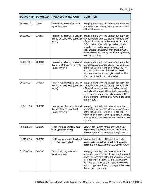
CONCEPTID<br />
399306005<br />
399239005<br />
399371001<br />
399036006<br />
399271003<br />
398998003<br />
399195005<br />
399310008<br />
<strong>SNOMED</strong>ID<br />
G-0397<br />
G-0398<br />
G-0399<br />
G-039A<br />
G-039B<br />
G-039C<br />
G-039D<br />
G-039E<br />
FULLY SPECIFIED NAME<br />
Parasternal short axis view<br />
(qualifier value)<br />
Parasternal short axis view at<br />
the aortic valve level (qualifier<br />
value)<br />
Parasternal short axis view at<br />
the level of the mitral chords<br />
(qualifier value)<br />
Parasternal short axis view at<br />
the mitral valve level (qualifier<br />
value)<br />
Parasternal short axis view at<br />
the papillary muscle level<br />
(qualifier value)<br />
Right ventricular inflow tract<br />
view (qualifier value)<br />
Right ventricular outflow tract<br />
view (qualifier value)<br />
Subcostal long axis view<br />
(qualifier value)<br />
DEFINITION<br />
<strong>Format</strong>s | 243<br />
Imaging plane with the transducer at the left<br />
sternal border oriented along the short axis<br />
of the left ventricle.<br />
Imaging plane with the transducer at the left<br />
sternal border oriented along the short axis<br />
of the left ventricle, at the base of the heart,<br />
IVC, atrial septum, tricuspid valve, which<br />
includes the aortic valve, right and left atria,<br />
right ventricular outflow tract and pulmonic<br />
valve, pulmonary artery, and in some patients,<br />
the LPA and RPA.<br />
Imaging plane with the transducer at the left<br />
sternal border oriented along the short axis<br />
of the left ventricle, which includes the left<br />
ventricle at the level of the mitral chords,<br />
ventricular septum, and right ventricle. This<br />
plane is inferior to the mitral valve.<br />
Imaging plane with the transducer at the left<br />
sternal border oriented along the short axis<br />
of the left ventricle, which includes the left<br />
ventricle at the level of the mitral valve leaflets,<br />
ventricular septum, and right ventricle. This<br />
plane is inferior to the aortic valve at the base<br />
of the heart.<br />
Imaging plane with the transducer at the<br />
sternal border oriented along the short axis<br />
of the left ventricle, which includes the left<br />
ventricle at the level of the papillary muscles,<br />
and right ventricle. This plane is inferior to the<br />
cordae.<br />
View of the Portion of the right ventricle<br />
adjacent to the tricuspid valve, the inflow<br />
portion of the RV. Common acronym: RVIT.<br />
View of the portion of the right ventricle<br />
adjacent to the pulmonic valve, the outflow<br />
portion of the RV. Common Acronym: RVOT.<br />
Imaging plane with the transducer at the<br />
subcostal space (inferior to sternum) oriented<br />
along the long axis of the left ventricle, which<br />
includes the left ventricle, left atrium, right<br />
ventricle and right atrium, septum between<br />
left and right ventricles, and septum between<br />
the left and right atria.<br />
© 2002-2012 International Health Terminology Standards Development Organisation CVR #: 30363434