Unit A Reproduction
Unit A Reproduction
Unit A Reproduction

You also want an ePaper? Increase the reach of your titles
YUMPU automatically turns print PDFs into web optimized ePapers that Google loves.
4.6<br />
Prenatal Procedures<br />
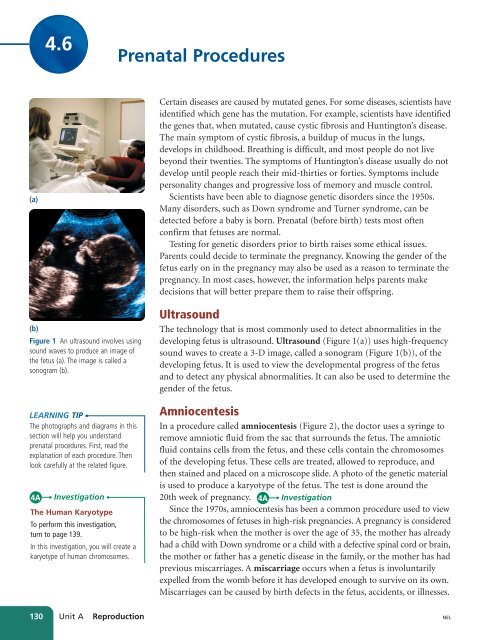
(a)<br />
(b)<br />
Figure 1 An ultrasound involves using<br />
sound waves to produce an image of<br />
the fetus (a). The image is called a<br />
sonogram (b).<br />
LEARNING TIP<br />
The photographs and diagrams in this<br />
section will help you understand<br />
prenatal procedures. First, read the<br />
explanation of each procedure. Then<br />
look carefully at the related figure.<br />
4A<br />
Investigation<br />
The Human Karyotype<br />
To perform this investigation,<br />
turn to page 139.<br />
In this investigation, you will create a<br />
karyotype of human chromosomes.<br />
Certain diseases are caused by mutated genes. For some diseases, scientists have<br />
identified which gene has the mutation. For example, scientists have identified<br />
the genes that, when mutated, cause cystic fibrosis and Huntington’s disease.<br />
The main symptom of cystic fibrosis, a buildup of mucus in the lungs,<br />
develops in childhood. Breathing is difficult, and most people do not live<br />
beyond their twenties. The symptoms of Huntington’s disease usually do not<br />
develop until people reach their mid-thirties or forties. Symptoms include<br />
personality changes and progressive loss of memory and muscle control.<br />
Scientists have been able to diagnose genetic disorders since the 1950s.<br />
Many disorders, such as Down syndrome and Turner syndrome, can be<br />
detected before a baby is born. Prenatal (before birth) tests most often<br />
confirm that fetuses are normal.<br />
Testing for genetic disorders prior to birth raises some ethical issues.<br />
Parents could decide to terminate the pregnancy. Knowing the gender of the<br />
fetus early on in the pregnancy may also be used as a reason to terminate the<br />
pregnancy. In most cases, however, the information helps parents make<br />
decisions that will better prepare them to raise their offspring.<br />
Ultrasound<br />
The technology that is most commonly used to detect abnormalities in the<br />
developing fetus is ultrasound. Ultrasound (Figure 1(a)) uses high-frequency<br />
sound waves to create a 3-D image, called a sonogram (Figure 1(b)), of the<br />
developing fetus. It is used to view the developmental progress of the fetus<br />
and to detect any physical abnormalities. It can also be used to determine the<br />
gender of the fetus.<br />
Amniocentesis<br />
In a procedure called amniocentesis (Figure 2), the doctor uses a syringe to<br />
remove amniotic fluid from the sac that surrounds the fetus. The amniotic<br />
fluid contains cells from the fetus, and these cells contain the chromosomes<br />
of the developing fetus. These cells are treated, allowed to reproduce, and<br />
then stained and placed on a microscope slide. A photo of the genetic material<br />
is used to produce a karyotype of the fetus. The test is done around the<br />
20th week of pregnancy. 4A Investigation<br />
Since the 1970s, amniocentesis has been a common procedure used to view<br />
the chromosomes of fetuses in high-risk pregnancies. A pregnancy is considered<br />
to be high-risk when the mother is over the age of 35, the mother has already<br />
had a child with Down syndrome or a child with a defective spinal cord or brain,<br />
the mother or father has a genetic disease in the family, or the mother has had<br />
previous miscarriages. A miscarriage occurs when a fetus is involuntarily<br />
expelled from the womb before it has developed enough to survive on its own.<br />
Miscarriages can be caused by birth defects in the fetus, accidents, or illnesses.<br />
130 <strong>Unit</strong> A <strong>Reproduction</strong><br />
NEL