Abstract Download (8.38MB)
Abstract Download (8.38MB)
Abstract Download (8.38MB)

You also want an ePaper? Increase the reach of your titles
YUMPU automatically turns print PDFs into web optimized ePapers that Google loves.
Name (Title):<br />
Keisuke Sato (MANA Research Associate)<br />
Affiliation:<br />
International Center for Materials Nanoarchitectonics<br />
(MANA), NIMS<br />
Address:<br />
1-1 Namiki, Tsukuba, Ibaraki 305-0044, Japan<br />
Email: SATO.Keisuke@nims.go.jp<br />
Home Page:<br />
Presentation Title:<br />
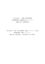
Silicon nanoparticles as fluorescent labeling of living cells<br />
<strong>Abstract</strong>:<br />
Fluorescent cadmium-based nanoparticles (NPs) have been<br />
developed as bio-imaging material in vitro and/or in vivo.<br />
However, concerns have been raised about the toxicological<br />
issue of cadmium in biological systems under ultraviolet (UV)exposure<br />
although the material has excellent optical properties,<br />
such as size-dependent tunable fluorescence color and high<br />
quantum yield of fluorescence for bio-imaging in vivo. Because<br />
of the problem, the development of new NPs consisting of nontoxic<br />
and more reliable elements is required. Silicon (Si)-NPs are<br />
candidate materials for the elimination of any potential<br />
toxicology problems. In this study, cytotoxicity of human<br />
cervical carcinoma cell line (HeLa) cells labeled with Si-NPs<br />
before and after UV-exposure, and the fluorescent imaging of Si-<br />
NPs in vivo is discussed. The cytotoxicity examined the viability<br />
of HeLa cells having Si-NPs as a function of the concentration<br />
using the mitochondrial activity assay. The UV-exposed Si-NPs<br />
exhibited a remarkably stable survival curve although the<br />
viability decreased only slight with increasing the concentration<br />
to 1.12 mg/ml, as shown in Fig. 1. This result substantiated the<br />
low toxicity of Si-NPs. Moreover, the fluorescent imaging using<br />
Si-NPs was investigated by the circulation from the lymphatic<br />
vessel to the lymph node of a mouse. The transfer pathway of Si-<br />
NPs could be satisfactorily recognized with the naked eye by<br />
detecting the strong red light from Si-NPs, as shown in Fig. 2.<br />
Therefore, these features demonstrate the usefulness of Si-NPs as<br />
the biological labels for living cell imaging in vitro and/or in<br />
vivo.<br />
References:<br />
[1] K. Sato, N. Kishimoto and K. Hirakuri, J. Appl. Phys. Vol.102 (2007) 104305.<br />
[2] K. Sato, N. Kishimoto and K. Hirakuri, J. Nanosci. Nanotechnol. Vol.8 (2008) 374.<br />
Viability ( % )<br />
100<br />
50<br />
0<br />
Unexposed<br />
UV-exposed<br />
Control<br />
10 -2<br />
Lymphatic vessel<br />
Poster Session PS-15<br />
10 -1<br />
10 0<br />
Concentration ( mg/ml )<br />
Fig. 1 Viability of HeLa cells having<br />
unexposed and UV-exposed Si-NPs<br />
as a function of the concentration.<br />
Lymph node<br />
Fig. 2 Fluorescence image of Si-NPs<br />
flowing into lymph node.<br />
85