Abstract Download (8.38MB)
Abstract Download (8.38MB)
Abstract Download (8.38MB)

You also want an ePaper? Increase the reach of your titles
YUMPU automatically turns print PDFs into web optimized ePapers that Google loves.
Name (Title):<br />
Yukio NAGASAKI (Professor)<br />
Affiliation:<br />
International Center for Materials Nanoarchitectonics Satellite (MANA),<br />
National Institute for Materials Science (NIMS) and Tsukuba Research<br />
Center for Interdisciplinary Materials Science(TIMS), University of Tsukuba<br />
Address:<br />
Tennoudai 1-1-1, Tsukuba, Ibaraki 305-8573, Japan.<br />
Email: Nagasaki@nagalabo.jp<br />
Home Page: http://www.ims.tsukuba.ac.jp/~nagasaki_lab/index.htm<br />
Presentation Title:<br />
Nano-particle Assisted Therapy<br />
<strong>Abstract</strong>:<br />
Nanosized polymeric particles have versatile utilities such as carrier for drug delivery,<br />
bioimaging and nano-diagnostic system. Recently, we have been focusing on preparation of<br />
core-shell type polymeric nanogels, which possess crosslinked polyamine core and biocompatible<br />
poly(ethylene glycol) (PEG) shell. The obtained nanogel showed volume phase transition in a<br />
minute change in environmental pH. Using the nanogel thus prepared as a nanoreactor, new<br />
nanomaterials were prepared for new therapy and imaging tools.<br />
Recently, we have found that gold nanoparticles (GNPs) were successfully synthesized<br />
through the self-reduction of Au(III) ions with PEGylated nanogels composed of polyamine gel<br />
core and PEG chains1. TEM images revealed that the synthesized GNPs located only in the<br />
nanogel core, indicating that nanogel acted as both nanoreactor and nanoreservoir for GNPs.<br />
Additionally, number of GNPs in sigle nanogel could be easily arranged by changing N / Au<br />
(number of amino groups / number of Au atoms) ratio. The obtained gold naoparticle containing<br />
nanogels (GNG) were applied for an enhancement of cancer phtothermal therapy as well as<br />
radiotherapy in response to electromagnetic wave in cultured cancer cells.<br />
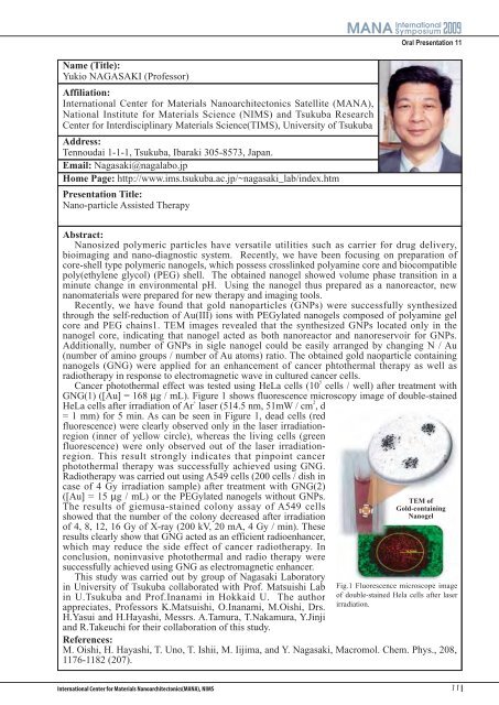
Cancer photothermal effect was tested using HeLa cells (10 5 cells / well) after treatment with<br />
GNG(1) ([Au] = 168 µg / mL). Figure 1 shows fluorescence microscopy image of double-stained<br />
HeLa cells after irradiation of Ar + laser (514.5 nm, 51mW / cm 2 , d<br />
= 1 mm) for 5 min. As can be seen in Figure 1, dead cells (red<br />
fluorescence) were clearly observed only in the laser irradiationregion<br />
(inner of yellow circle), whereas the living cells (green<br />
fluorescence) were only observed out of the laser irradiationregion.<br />
This result strongly indicates that pinpoint cancer<br />
photothermal therapy was successfully achieved using GNG.<br />
Radiotherapy was carried out using A549 cells (200 cells / dish in<br />
case of 4 Gy irradiation sample) after treatment with GNG(2)<br />
([Au] = 15 µg / mL) or the PEGylated nanogels without GNPs.<br />
The results of giemusa-stained colony assay of A549 cells<br />
showed that the number of the colony decreased after irradiation<br />
of 4, 8, 12, 16 Gy of X-ray (200 kV, 20 mA, 4 Gy / min). These<br />
results clearly show that GNG acted as an efficient radioenhancer,<br />
which may reduce the side effect of cancer radiotherapy. In<br />
conclusion, noninvasive photothermal and radio therapy were<br />
successfully achieved using GNG as electromagnetic enhancer.<br />
This study was carried out by group of Nagasaki Laboratory<br />
in University of Tsukuba collaborated with Prof. Matsuishi Lab<br />
in U.Tsukuba and Prof.Inanami in Hokkaid U. The author<br />
appreciates, Professors K.Matsuishi, O.Inanami, M.Oishi, Drs.<br />
H.Yasui and H.Hayashi, Messrs. A.Tamura, T.Nakamura, Y.Jinji<br />
and R.Takeuchi for their collaboration of this study.<br />
References:<br />
Oral Presentation 11<br />
TEM of<br />
Gold-containing<br />
Nanogel<br />
Fig.1 Fluorescence microscope image<br />
of double-stained Hela cells after laser<br />
irradiation.<br />
M. Oishi, H. Hayashi, T. Uno, T. Ishii, M. Iijima, and Y. Nagasaki, Macromol. Chem. Phys., 208,<br />
1176-1182 (207).<br />
11