Abstract Download (8.38MB)
Abstract Download (8.38MB)
Abstract Download (8.38MB)

Create successful ePaper yourself
Turn your PDF publications into a flip-book with our unique Google optimized e-Paper software.
Name (Title):<br />
Kohsaku Kawakami (MANA Researcher, Senior Researcher)<br />
Affiliation:<br />
International Center for Materials Nanoarchitectonics<br />
(MANA) and Biomaterials Center, NIMS<br />
Address:<br />
1-1 Namiki, Tsukuba. Ibaraki 305-0044, Japan<br />
Email: KAWAKAMI.Kohsaku@nims.go.jp<br />
Home Page: http://www.nims.go.jp/bmc/group/ndg/kawakami/<br />
Presentation Title:<br />
Electrospray Deposition as a New Method for Preparing Fine Drug Particles<br />
Poster Session PB-3<br />
<strong>Abstract</strong>:<br />
Although there has been much discussion on favorable physicochemical properties for drug<br />
candidates over the last decade, formulators must still deal with many challenging compounds<br />
including poorly soluble drugs. A novel formulation technology, electrospray deposition method,<br />
is introduced in this presentation. This method enabled amorphization and micronization of the<br />
formulation, both of which contribute to improvement of the solubility, in one-step preparation<br />
procedure.<br />
Electrospray Deposition (called as “Electrospinning” in polymer science field) has been<br />
regarded as a promising method to prepare various fibrous materials such as filters, sensors, and<br />
many types of biomedical materials. Various poorly soluble drugs including predonisolone<br />
(PDN), phenylbutazone, phenytoin and carbamazepine were dissolved with polymeric carriers<br />
including polyvinylpyrroridone (PVP), Eudragid E-100, and polyethylene glycol either in ethanol<br />
or in its aqueous solution. The solutions were introduced to 8-pin manifold using a syringe pump,<br />
followed by application of high voltage, typically in the range of 10-25 kV, to produce<br />
electrospray to a stainless steel target. The setup was enclosed in an air- tight box to control<br />
humidity of the environment. The dried particles were collected from the target surface.<br />
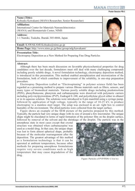
Figure shows an example of the SEM picture of the particles prepared by this method.<br />
Typically, the particle had very unique shape with its size of around a few micrometer. The unique<br />
shape might be elucidated in terms of rapid formation of the polymer film on the droplet surface,<br />
followed by removal of the solvent and the shrinkage of the droplet. The particle was in the<br />
amorphous state in most cases except that crystal<br />
structure was obtained when carbamazepine was<br />
used as a model drug. In that case, the unique shape<br />
was lost to form almost spherical shape, probably<br />
because of the molecular rearrangement after the<br />
deposition. The greatest advantage of this method<br />
as a formulation technology is that it can be<br />
operated at ambient temperature, because other<br />
methods for preparing amorphous formulations<br />
require very severe conditions such as high<br />
temperature that may cause degradation of the<br />
drug.<br />
Figure: Particles of PVP/PDN=1/1(w/w)<br />
97