Physical Principles of Electron Microscopy: An Introduction to TEM ...
Physical Principles of Electron Microscopy: An Introduction to TEM ...
Physical Principles of Electron Microscopy: An Introduction to TEM ...

Create successful ePaper yourself
Turn your PDF publications into a flip-book with our unique Google optimized e-Paper software.
The Scanning <strong>Electron</strong> Microscope 137<br />
5.4 Backscattered-<strong>Electron</strong> Images<br />
A backscattered electron (BSE) is a primary electron that has been ejected<br />
from a solid by scattering through an angle greater than 90 degrees. Such<br />
deflection could occur as a result <strong>of</strong> several collisions, some or all <strong>of</strong> which<br />
might involve a scattering angle <strong>of</strong> less than 90 degrees; however, a single<br />
elastic event with � > 90 degrees is quite probable. Because the elastic<br />
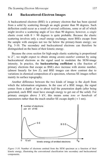
scattering involves only a small energy exchange, most BSEs escape from<br />
the sample with energies not <strong>to</strong>o far below the primary-beam energy; see<br />
Fig. 5-10. The secondary and backscattered electrons can therefore be<br />
distinguished on the basis <strong>of</strong> their kinetic energy.<br />
Because the cross section for high-angle elastic scattering is proportional<br />
<strong>to</strong> Z 2 , we might expect <strong>to</strong> obtain strong a<strong>to</strong>mic-number contrast by using<br />
backscattered electrons as the signal used <strong>to</strong> modulate the SEM-image<br />
intensity. In practice, the backscattering coefficient � (the fraction <strong>of</strong><br />
primary electrons that escape as BSE) does increase with a<strong>to</strong>mic number,<br />
(almost linearly for low Z), and BSE images can show contrast due <strong>to</strong><br />
variations in chemical composition <strong>of</strong> a specimen, whereas SE images reflect<br />
mainly its surface <strong>to</strong>pography.<br />
<strong>An</strong>other difference between the two kinds <strong>of</strong> image is the depth from<br />
which the information originates. In the case <strong>of</strong> a BSE image, the signal<br />
comes from a depth <strong>of</strong> up <strong>to</strong> about half the penetration depth (after being<br />
generated, each BSE must have enough energy <strong>to</strong> get out <strong>of</strong> the solid). For<br />
primary energies above 3 kV, this means some tens or hundreds <strong>of</strong><br />
nanometers rather than the much smaller SE escape depth ( � 1 nm).<br />
0<br />
number <strong>of</strong> electrons<br />
per eV <strong>of</strong> KE<br />
10 eV<br />
S E<br />
B S E<br />
kinetic energy <strong>of</strong> emitted electrons<br />
Figure 5-10. Number <strong>of</strong> electrons emitted from the SEM specimen as a function <strong>of</strong> their<br />
kinetic energy, illustrating the conventional classification in<strong>to</strong> secondary and backscattered<br />
components.<br />
E 0