Physical Principles of Electron Microscopy: An Introduction to TEM ...
Physical Principles of Electron Microscopy: An Introduction to TEM ...
Physical Principles of Electron Microscopy: An Introduction to TEM ...

You also want an ePaper? Increase the reach of your titles
YUMPU automatically turns print PDFs into web optimized ePapers that Google loves.
<strong>An</strong> <strong>Introduction</strong> <strong>to</strong> <strong>Microscopy</strong> 15<br />
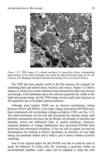
Figure 1-12. <strong>TEM</strong> image <strong>of</strong> a stained specimen <strong>of</strong> mouse-liver tissue, corresponding<br />
approximately <strong>to</strong> the small rectangular area within the light-microscope image on the left.<br />
Courtesy <strong>of</strong> R. Bhatnagar, Biological Sciences <strong>Microscopy</strong> Unit, University <strong>of</strong> Alberta.<br />
The <strong>TEM</strong> has been equally useful in the life sciences, for example for<br />
examining plant and animal tissue, bacteria, and viruses. Figure 1-12 shows<br />
images <strong>of</strong> mouse-liver tissue obtained using transmission light and electron<br />
microscopes. Cell membranes and a few internal organelles are visible in the<br />
light-microscope image, but the <strong>TEM</strong> image reveals much more structure in<br />
the organelles, due <strong>to</strong> its higher spatial resolution.<br />
Although most modern <strong>TEM</strong>s use an electron accelerating voltage<br />
between 100 kV and 300 kV, a few high-voltage instruments (HVEMs) have<br />
been constructed with accelerating voltages as high as 3 MV; see Fig. 1-13.<br />
The main motivation was the fact that increasing the electron energy (and<br />
therefore momentum) decreases the de Broglie wavelength <strong>of</strong> electrons and<br />
therefore lowers the diffraction limit <strong>to</strong> spatial resolution. However,<br />
technical problems <strong>of</strong> voltage stabilization have prevented HVEMs from<br />
achieving their theoretical resolution. A few are still in regular use and are<br />
advantageous for looking at thicker specimens, as electrons <strong>of</strong> very high<br />
energy can penetrate further in<strong>to</strong> a solid (1 �m or more) without excessive<br />
scattering.<br />
One <strong>of</strong> the original hopes for the HVEM was that it could be used <strong>to</strong><br />
study the behavior <strong>of</strong> living cells. By enclosing a specimen within an<br />
environmental chamber, water vapor can be supplied <strong>to</strong> keep the cells