Physical Principles of Electron Microscopy: An Introduction to TEM ...
Physical Principles of Electron Microscopy: An Introduction to TEM ...
Physical Principles of Electron Microscopy: An Introduction to TEM ...

Create successful ePaper yourself
Turn your PDF publications into a flip-book with our unique Google optimized e-Paper software.
The Scanning <strong>Electron</strong> Microscope 145<br />
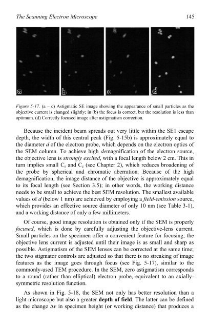
Figure 5-17. (a – c) Astigmatic SE image showing the appearance <strong>of</strong> small particles as the<br />
objective current is changed slightly; in (b) the focus is correct, but the resolution is less than<br />
optimum. (d) Correctly focused image after astigmatism correction.<br />
Because the incident beam spreads out very little within the SE1 escape<br />
depth, the width <strong>of</strong> this central peak (Fig. 5-15b) is approximately equal <strong>to</strong><br />
the diameter d <strong>of</strong> the electron probe, which depends on the electron optics <strong>of</strong><br />
the SEM column. To achieve high demagnification <strong>of</strong> the electron source,<br />
the objective lens is strongly excited, with a focal length below 2 cm. This in<br />
turn implies small Cs and Cc (see Chapter 2), which reduces broadening <strong>of</strong><br />
the probe by spherical and chromatic aberration. Because <strong>of</strong> the high<br />
demagnification, the image distance <strong>of</strong> the objective is approximately equal<br />
<strong>to</strong> its focal length (see Section 3.5); in other words, the working distance<br />
needs <strong>to</strong> be small <strong>to</strong> achieve the best SEM resolution. The smallest available<br />
values <strong>of</strong> d (below 1 nm) are achieved by employing a field-emission source,<br />
which provides an effective source diameter <strong>of</strong> only 10 nm (see Table 3-1),<br />
and a working distance <strong>of</strong> only a few millimeters.<br />
Of course, good image resolution is obtained only if the SEM is properly<br />
focused, which is done by carefully adjusting the objective-lens current.<br />
Small particles on the specimen <strong>of</strong>fer a convenient feature for focusing; the<br />
objective lens current is adjusted until their image is as small and sharp as<br />
possible. Astigmatism <strong>of</strong> the SEM lenses can be corrected at the same time;<br />
the two stigma<strong>to</strong>r controls are adjusted so that there is no streaking <strong>of</strong> image<br />
features as the image goes through focus (see Fig. 5-17), similar <strong>to</strong> the<br />
commonly-used <strong>TEM</strong> procedure. In the SEM, zero astigmatism corresponds<br />
<strong>to</strong> a round (rather than elliptical) electron probe, equivalent <strong>to</strong> an axiallysymmetric<br />
resolution function.<br />
As shown in Fig. 5-18, the SEM not only has better resolution than a<br />
light microscope but also a greater depth <strong>of</strong> field. The latter can be defined<br />
as the change �v in specimen height (or working distance) that produces a