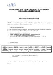
4(%3)3 - Ecole nationale supérieure de chimie de Montpellier
4(%3)3 - Ecole nationale supérieure de chimie de Montpellier
4(%3)3 - Ecole nationale supérieure de chimie de Montpellier

Create successful ePaper yourself
Turn your PDF publications into a flip-book with our unique Google optimized e-Paper software.
To prepare the monolayer assembly of the micelles, first a substrate layer of uncontrolledPMMA was spin coated over thoroughly cleaned Silicon wafer using spin coating. 1 g PMMA wasdissolved in 3 ml Dicholoromethane in a small vial and the resulting solution was coated onto theSilicon wafer dried un<strong>de</strong>r argon atmosphere in the spin coater. The spin rate was kept at 1000 rpmfor 3 minutes to obtain 1.3 µm thick substrate film. The coated substrate thus obtained wasimmersed in small beaker a 10 ml micellar solution of PMMA-b-PODMA (3.5 g.ml -1 inTHF/Cyclohexane, 1/9 vol.%) for 1 hour followed by rinsing with Cyclohexane. The coated surfacethus obtained was dried and further analyzed.To prepare multilayer assembly, the PMMA coated wafer was first swelled in 20 ml mixtureof THF/Cyclohexane (1/9, vol.%) for 15 minutes. The swelled PMMA coated wafer was taken outquickly, put into spin coater and the micellar solution of PMMA-b-PODMA (3.5 g.ml -1 ) was coatedun<strong>de</strong>r argon atmosphere.8.3.7 Contact Angle MeasurementsThe contact angle measurements were conducted by drop sessile methond on lab ma<strong>de</strong>apparatus equipped with a light source, a stage to put the substrate to be analyzed, a light sourceand a CCD camera. A 10 µl water drop was dropped onto the sample surface by using a 50 µl syringestationed above the stage. All measurements were done at room temperature and the imagesacquired were analyzed using ImageJ® software and the contact angles were <strong>de</strong>termined byinterpolation method using DropSnake® plugin. The values reported represent average of 5measurements.8.3.8 Atomic Force MicroscopyThe AFM instrumentation and its specifications remains the same as mentioned in the firstsection of this chapter.8.3.9 Scanning Electron MicroscopyThe SEM instrumentation and its specifications are the same as mentioned in the firstsection. The samples were metalized for 6 minutes before the analysis.8.4 NANO-GEL BASED SELF-HEALING SYSTEM152