

View PDF Version - RePub - Erasmus Universiteit Rotterdam
View PDF Version - RePub - Erasmus Universiteit Rotterdam
View PDF Version - RePub - Erasmus Universiteit Rotterdam

You also want an ePaper? Increase the reach of your titles
YUMPU automatically turns print PDFs into web optimized ePapers that Google loves.
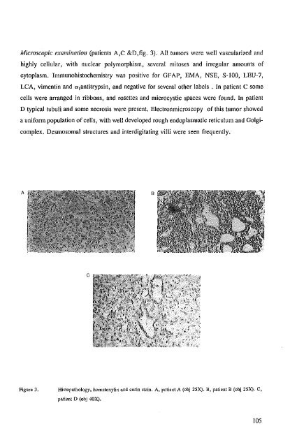
Microscopic examination (patients A,C &D,fig. 3). All tumors were well vascularized and<br />
highly cellular, with nuclear polymorphism, several mitoses and irregular amounts of<br />
cytoplasm. Immunohistochemistry was positive for GFAP, EMA, NSE, S-100, LEU-?,<br />
LCA, vimentin and 0i1antitrypsin, and negative for several other labels. In patient C some<br />
cells were arranged in ribbons, and rosettes and microcystic spaces were found. In patient<br />
D typical tubuli and some necrosis were present. Electronmicroscopy of this tumor showed<br />
a uniform population of cells, with well developed rough endoplasmatic reticulum and Golgicomplex.<br />
Desmosomal structures and interdigitating villi were seen frequently.<br />
A<br />
B<br />
c<br />
Figure 3,<br />
Histopathology, hematoxylin and eosin stain. A, patient A (obj 25X), B, patient B (obj 25X). C,<br />
patient D (obj 40X).<br />
105


