

View PDF Version - RePub - Erasmus Universiteit Rotterdam
View PDF Version - RePub - Erasmus Universiteit Rotterdam
View PDF Version - RePub - Erasmus Universiteit Rotterdam

Create successful ePaper yourself
Turn your PDF publications into a flip-book with our unique Google optimized e-Paper software.
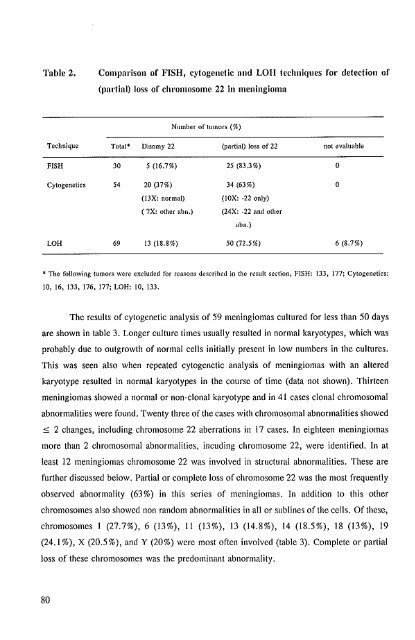
Table 2.<br />
Comparison of FISH, cytogenetic and LOn techniqnes for detection of<br />
(paltinl) loss of chl'omosome 22 in meningioma<br />
Number of tumors (%)<br />
Technique<br />
FISH<br />
Cytogenetics<br />
LOH<br />
Total* Disomy 22 (partial) loss of 22 not evaluable<br />
30 5 (16.7%) 25 (83.3%) o<br />
54 20 (37%) 34 (63%) o<br />
(13X: normal)<br />
(lOX: -22 only)<br />
( 7X: other abn.) (24X: -22 and other<br />
aim.)<br />
69 13 (18.8%) 50 (72.5%) 6 (8.7%)<br />
* The following tumors were excluded for rea~ons described in the result section, FISH: 1:33, 177; Cytogenetics:<br />
10, 16, 133, 176. 177; LOH: 10, 133.<br />
The results of cytogenelic analysis of 59 meningiomas cultured for less than 50 days<br />
are shown in table 3. Longer culture times usually resulted in normal karyotypes, which was<br />
probably due to outgrowth of normal cells initially present in low numbers in the cultures.<br />
This was seen also when repeated cytogenetic analysis of meningiomas with an altered<br />
karyotype resulted in normal karyotypes in the course of time (data nol shown). Thirteen<br />
meningiomas showed a normal or non-clonal karyotype and in 41 cases clonal chromosomal<br />
abnormalities were found. Twenty three of the cases with chromosomal abnormalities showed<br />
::;; 2 changes, including chromosome 22 aberrations in 17 cases. In eighteen meningiomas<br />
more than 2 chromosomal abnormalities, incuding chromosome 22, were identified. In at<br />
least 12 meningiomas chromosome 22 was involved in structural abnormalities. These are<br />
further discussed below. Partial or complete loss of chromosome 22 was the most frequently<br />
observed abnormality (63%) in this series of meningiomas. In addition to this other<br />
chromosomes also showed non random abnormalities in all or sublines of the cells. Of these,<br />
chromosomes I (27.7%), 6 (13%), II (13%), 13 (14.8%), 14 (18.5%), 18 (13%), 19<br />
(24.1 %), X (20.5%), and Y (20%) were most of len involved (table 3). Complete or partial<br />
loss of these chromosomes was the predominant abnormality.<br />
80


