

View PDF Version - RePub - Erasmus Universiteit Rotterdam
View PDF Version - RePub - Erasmus Universiteit Rotterdam
View PDF Version - RePub - Erasmus Universiteit Rotterdam

Create successful ePaper yourself
Turn your PDF publications into a flip-book with our unique Google optimized e-Paper software.
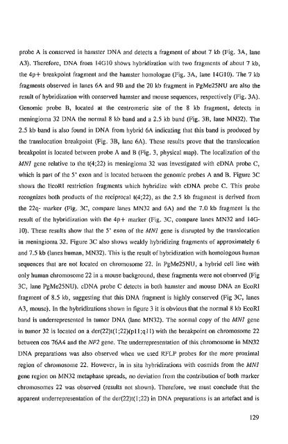
probe A is conserved in hamster DNA and detects a fragment of about 7 kb (Fig. 3A, lane<br />
A3). Therefore, DNA from 14010 shows hybridization with two fragments of about 7 kb,<br />
the 4p+ breakpoint fragment and the hamster homologue (Fig. 3A, lane 14010). The 7 kb<br />
fragments observed in lanes 6A and 9B and the 20 kb fragment in PgMe25NU are also the<br />
result of hybridization with conserved hamster and mouse sequences, respectively (Fig. 3A).<br />
Oenomic probe B, located at the centromeric site of the 8 kb fragment, detects in<br />
meningioma 32 DNA the normal 8 kb band and a 2.5 kb band (Fig. 3B, lane MN32). The<br />
2.5 kb band is also found in DNA from hybrid 6A indicating that this band is produced by<br />
the translocation breakpoint (Fig. 3B, lane 6A). These results prove that the translocation<br />
breakpoint is located between probe A and B (Fig. 3, physical map). The localization of the<br />
MNl gene relative to the t(4;22) in meningioma 32 was investigated with cDNA probe C,<br />
which is part of the 5' exon and is located between the genomic probes A and B. Figure 3C<br />
shows the EcoRI restriction fragments which hybridize with cDNA probe C. This probe<br />
recognizes both products of the reciprocal t(4;22), as the 2.5 kb fragment is derived from<br />
the 22q- marker (Fig. 3C, compare lanes MN32 and 6A) and the 7.0 kb fragment is the<br />
result of the hybridization with the 4p+ marker (Fig. 3C, compare lanes MN32 and 140-<br />
10). These results show that the 5' exon of the MNI gene is disrupted by the translocation<br />
in meningioma 32. Figure 3C also shows weakly hybridizing fragments of approximately 6<br />
and 7.5 kb (lanes human, MN32). This is the result of hybridization with homologous human<br />
sequences that are not located on chromosome 22. In PgMe25NU, a hybrid ceHline with<br />
only human chromosome 22 in a mouse background, these fragments were not observed (Fig<br />
3C, lane PgMe25NU). cDNA probe C detects in both hamster and mouse DNA an EcoRI<br />
fragment of 8.5 kb, suggesting that this DNA fragment is highly conserved (Fig 3C, lanes<br />
A3, mouse). In the hybridizations shown in figure 3 it is obvious that the normal 8 kb EcoRI<br />
band is underrepresented in tumor DNA (lane MN32). The normal copy of the MNI gene<br />
in tumor 32 is located on a der(22)t( I ;22)(p II ;q II) with the breakpoint on chromosome 22<br />
between cos 76A4 and the NF2 gene. The underrepresentation of this chromosome in MN32<br />
DNA preparations was also observed when we used RFLP probes for the more proximal<br />
region of chromosome 22. However, in in situ hybridizations with cosmids from the MNI<br />
gene region on MN32 metaphase spreads, no deviation from the contribution of both marker<br />
chromosomes 22 was observed (results not shown). Therefore, we must conclude that the<br />
apparent underrepresentation of the der(22)t( I ;22) in DNA preparations is an artefact and is<br />
129


