

View PDF Version - RePub - Erasmus Universiteit Rotterdam
View PDF Version - RePub - Erasmus Universiteit Rotterdam
View PDF Version - RePub - Erasmus Universiteit Rotterdam

Create successful ePaper yourself
Turn your PDF publications into a flip-book with our unique Google optimized e-Paper software.
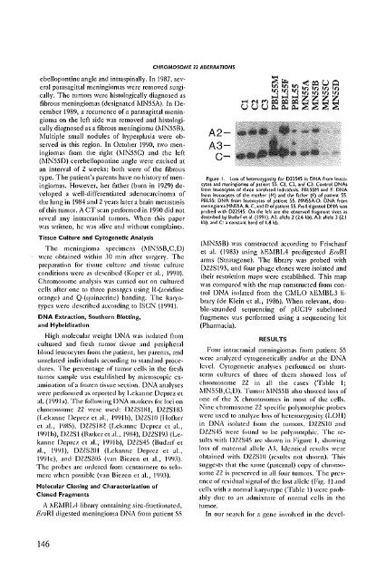
CHROMOSOME 11 ABERRATIONS<br />
ebellopontine angle and intraspinally. In 1987, several<br />
pamsagittal meningiomas were removed surgically.<br />
The tumors were histologically diagnosed as<br />
fibrous meningiomas (designated ~[N55A). In December<br />
1989, a reCLlrrence of a parasagittal meningioma<br />
on the left side was removed and histologically<br />
diagnosed as a fibrous meningioma (:..tN55B).<br />
Multiple small nodules of hyperplasia were observed<br />
in this region. In October 19l)O, two meningiomas<br />
from the right (:"IN55C) and the left<br />
(i\IN550) cerebellopolltine angle were excised at<br />
an inten'al of 2 weeks; both were of the fibrous<br />
type. The patient's parents have no history of meningiomas.<br />
However, her t~lther (born in 1929) developed<br />
a well-differentiated adenocarcinoma of<br />
the lung in 19H4 and 2 years latera brain metastasis<br />
of this tumor. A CT scan performed in 1990 did not<br />
reveal any intracmnial tumors. When this paper<br />
W,IS written, he was alive ilntl without complaints.<br />
Tissue Culture and Cytogenetic Analysis<br />
The meningioma specimens (MN55B,C,D)<br />
were ohtained within 3{) min after surgery. The<br />
preparation for tissue culture and tissue culture<br />
l'Onditions were as described (Koper et aI., 1990).<br />
Chromosome analysis WilS carried Ollt on cultured<br />
cells after one to three passages using R-(acridine<br />
omnge) and Q-(quinacrine) banding. The karyotypes<br />
were described aCl'onling to ISeN (1991).<br />
DNA Extraction, Southern Blotting,<br />
and Hybridization<br />
High molecular weight DNA was isolated from<br />
I:ultured and fresh tumor ti.~sue and peripheral<br />
blood leucocytes from the patient, her parents, and<br />
unrelated individuals according tu standard procedures.<br />
The percentage of tumor cells in the fresh<br />
tumor sample was est,lhlished by microscopic examination<br />
of a frozen tissue section. DNA analyses<br />
were performed as reported by Lekanne Deprez e(<br />
a!. (199b). The following DNA markers for loci on<br />
chromosome 22 were used: D2ZSIHI, D2ZS183<br />
(Lekanne Deprez et aI., 1


