View PDF Version - RePub - Erasmus Universiteit Rotterdam
View PDF Version - RePub - Erasmus Universiteit Rotterdam
View PDF Version - RePub - Erasmus Universiteit Rotterdam
You also want an ePaper? Increase the reach of your titles
YUMPU automatically turns print PDFs into web optimized ePapers that Google loves.
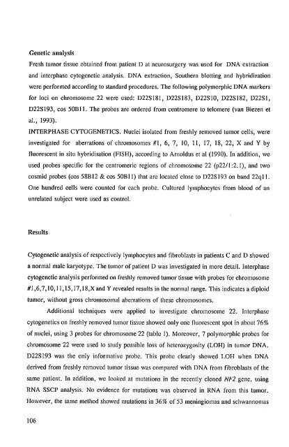
Genetic analysis<br />
Fresh tumor tissue obtained from patient D at neurosurgery was used for DNA extraction<br />
and interphase cytogenetic analysis. DNA extraction, Southern blotting and hybridization<br />
were performed according 10 standard procedures. The following polymorphic DNA markers<br />
for loci on chromosome 22 were used: D22S181, D22S183, D22SIO, D22S182, D22SI,<br />
D22S 193, cos 50BII. The probes are ordered from centromere to telomere (van Biezen et<br />
aI., 1993).<br />
INTERPHASE CYTOGENETICS. Nuclei isolated from freshly removed tumor cells, were<br />
investigated for aberrations of chromosomes #1, 6, 7, 10, 11, 17, 18, 22, X and Y by<br />
fluorescent in situ hybridisation (FISH), according to Arnoldus et al (1990). In addition, we<br />
used probes specific for the centromeric regions of chromosome 22 (p22!1 :2.1), and two<br />
cosmid probes (cos 58BI2 & cos 50BII) that are located close to D22S193 on band 22qll.<br />
One hundred cells were counted for each probe. Cultured lymphocytes from blood of an<br />
unrelated subject were used as control.<br />
Results<br />
Cytogenetic analysis of respectively lymphocytes and fibroblasts in patients C and D showed<br />
a normal male karyotype. The tumor of patient D was investigated in more detail. Interphase<br />
cytogenetic analysis performed on freshly removed tumor tissue with probes for chromosome<br />
#1,6,7,10,11, IS, 17, 18,X and Y revealed results in the normal range. This indicates a diploid<br />
tumor, without gross chromosomal aberrations of these chromosomes.<br />
Additional techniques were applied to investigate chromosome 22. Interphase<br />
cytogenetics on freshly removed tumor tissue showed only one tluorescent spot in about 76%<br />
of nuclei, using 3 probes for chromosome 22 (table I). Moreover, 7 polymorphic probes for<br />
chromosome 22 were used to study possible loss of heterozygosity (LOH) in tumor DNA.<br />
D22S193 was the only informative probe. This probe clearly showed LOH when DNA<br />
derived from freshly removed tumor tissue was compared with DNA from fibroblasts of the<br />
same patient. In addition, we looked at mutations in the recently cloned NF2 gene, using<br />
RNA SSCP analysis. No evidence for mutations was observed in RNA from this tumor.<br />
However, the same method showed mutations in 36% of 53 meningiomas and schwannomas<br />
106


