

View PDF Version - RePub - Erasmus Universiteit Rotterdam
View PDF Version - RePub - Erasmus Universiteit Rotterdam
View PDF Version - RePub - Erasmus Universiteit Rotterdam

You also want an ePaper? Increase the reach of your titles
YUMPU automatically turns print PDFs into web optimized ePapers that Google loves.
(Lekanne Deprez et aI., 1994).<br />
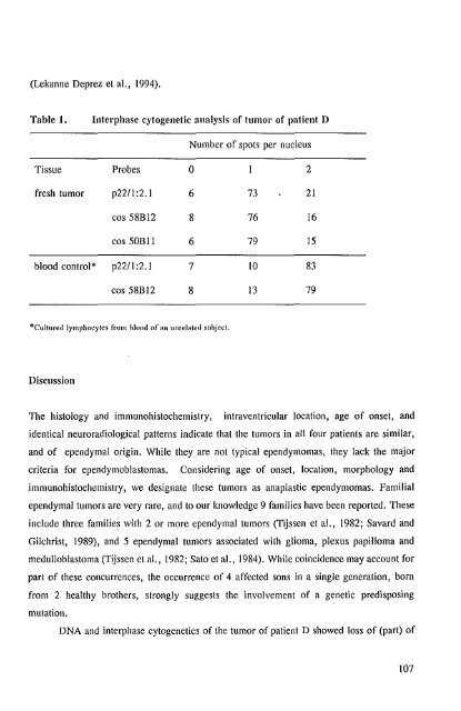
Table 1.<br />
Interphase cytogenetic analysis of tumor of patient D<br />
Number of spots per nucleus<br />
Tissue Probes 0 2<br />
fresh tumor p221l :2.1 6 73 21<br />
cos 58BI2 8 76 16<br />
cos 50BII 6 79 15<br />
blood control* p221l :2.1 7 IO 83<br />
cos 58BI2 8 13 79<br />
*Cultured lymphocytes from blood of an unrelated subject.<br />
Discussion<br />
The histology and immunohistochemistry, intraventricular location, age of onset, and<br />
identical neuroradiological patterns indicate that the tumors in all four patients are similar,<br />
and of ependymal origin. While they are not typical ependymomas, they lack the major<br />
criteria for ependymoblastomas. Considering age of onset, location, morphology and<br />
immunohistochemistry, we designate these tumors as anaplastic ependymomas. Familial<br />
ependymal tumors are very rare, and to our knowledge 9 families have been reported. These<br />
include three families with 2 or more ependymal tumors (Tijssen et aI., 1982; Savard and<br />
Gilchrist, 1989), and 5 ependymal tumors associated with glioma, plexus papilloma and<br />
medulloblastoma (Tijssen et aI., 1982; Sato et aI., 1984). While coincidence may account for<br />
part of these concurrences, the occurrence of 4 affected sons in a single generation, born<br />
from 2 healthy brothers, strongly suggests the involvement of a genetic predisposing<br />
mutation.<br />
DNA and interphase cytogenetics of the tumor of patient D showed loss of (part) of<br />
107


