- Page 1:
Slovenská spoločnosť pre bioché
- Page 4 and 5:
XXII. Biochemistry Congress is supp
- Page 6 and 7:
Wednesday, 8 September 2010 14:00 -
- Page 8 and 9:
Program in details: Wednesday PROGr
- Page 10 and 11:
Program in details: Thursday 11.35
- Page 12 and 13:
Program in details: Thursday 15.45
- Page 14 and 15:
Program in details: Friday friday,
- Page 16 and 17:
Program in details: Friday Jesseniu
- Page 18 and 19:
Program in details: Saturday SATURD
- Page 20 and 21:
Program in details: Saturday Jessen
- Page 22 and 23:
Program in details: Sunday SUNDAY,
- Page 24 and 25:
Program in details: Poster viewing
- Page 26 and 27:
Program in details: Poster viewing
- Page 28 and 29:
Program in details: Poster viewing
- Page 30 and 31:
Program in details: Poster viewing
- Page 32 and 33:
Program in details: Poster viewing
- Page 34 and 35:
Program in details: Poster viewing
- Page 36 and 37:
34 XXII. Biochemistry Congress, Mar
- Page 38 and 39:
Plenary lectures IDENTIFICATION OF
- Page 40 and 41:
Plenary lectures STRUCTUraL-FUNCTIO
- Page 42 and 43:
LECTURES 40 XXII. Biochemistry Cong
- Page 44 and 45:
Lectures LIPID HELICES formaTION IN
- Page 46 and 47:
Lectures SYNTHESIS OF GLCNaC-TS MIM
- Page 48 and 49:
Lectures TOXCAT METHOD: APPLICATION
- Page 50 and 51:
Lectures PROTEOMICS OF MULTIFUNCTIO
- Page 52 and 53:
Lectures BIOMarKErs of LYMPH NODE M
- Page 54 and 55:
Lectures OSTERIX OVER-EXPRESSION IN
- Page 56 and 57: Lectures MAGNETIC RESONANCE SPECTRO
- Page 58 and 59: Lectures TraNSGLYCOSYLATION - a UNI
- Page 60 and 61: Lectures TWENTY fOUr YEars SINCE CH
- Page 62 and 63: Lectures SPECIALITIES OF OXIDATIVE
- Page 64 and 65: Lectures DO WE TEACH BIOCHEMISTRY I
- Page 66 and 67: Lectures BRONCHIAL ASTHMA AND EffEC
- Page 68 and 69: Lectures NEW POSSIBILITIES FOR THE
- Page 70 and 71: Lectures MOLECULar MECHaNISMS INvOL
- Page 72 and 73: Lectures TEACHING BIOCHEMISTRY AT T
- Page 74 and 75: Lectures Assembly of BaCILLus suBTI
- Page 76 and 77: Lectures GATING OF THE T-TYPE CALCI
- Page 78 and 79: Lectures SPINAL CORD INJURY: PATHOG
- Page 80 and 81: Lectures BIOCHEMISTry IN THE PICTUr
- Page 82 and 83: Lectures THE IDENTIfICaTION aND CHa
- Page 84 and 85: Lectures RTX CYTOTOXINS rECOGNIZE
- Page 86 and 87: Lectures oxidaTIve rISK IN aTHErOSC
- Page 88 and 89: Lectures AcrIDINES - USEfUL MUTaGEN
- Page 90 and 91: Lectures INTrONIC LINE-1 INSErTION
- Page 92 and 93: Lectures Is PHOSPHaTIDYLINOSITOL tr
- Page 94 and 95: Lectures E-LEarNING - FrIEND of fOE
- Page 96 and 97: Lectures XENOBIOTIC-METABOLIZING EN
- Page 98 and 99: Lectures ENErGETIC aSPECTS of a MOD
- Page 100 and 101: Lectures CYTOCHrOME P450- aND PErOX
- Page 102 and 103: Lectures MYCOBaCTErial maNNOSYL tra
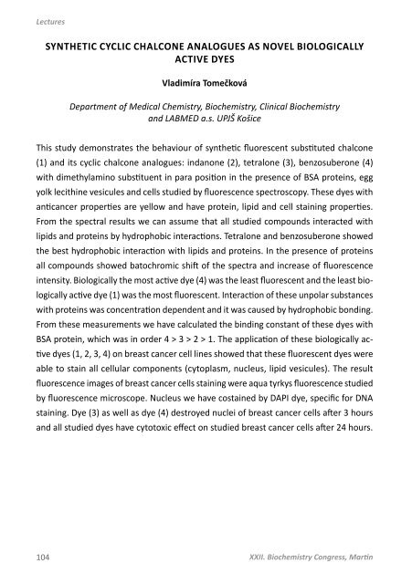
- Page 104 and 105: Lectures WHEN MOre IS LESS: a DILEM
- Page 108 and 109: Lectures LivING COLOUrs: ILLUMINaTE
- Page 110 and 111: Lectures ATOrvaSTATIN CHANGES MEMBr
- Page 112 and 113: Lectures LECTINS frOM PATHOGENS: MY
- Page 114 and 115: Lectures THE ROLE OF ANGIOTENSIN II
- Page 116 and 117: Posters I. BIOCHEMISTry aND MOLECUL
- Page 118 and 119: Posters 2. IS IT POSSIBLE TO IMPROV
- Page 120 and 121: Posters 4. An anaLYSIS of THE IMPaC
- Page 122 and 123: Posters 6. ANATOMICAL DISTRIBUTION
- Page 124 and 125: Posters II. BIOTECHNOLOGY 122 XXII.
- Page 126 and 127: Posters 9. EffECT of METHYL jaSMONa
- Page 128 and 129: Posters 11. DESIGN of an EXPrESSION
- Page 130 and 131: Posters 13. EXPRESSION, PURIFICATIO
- Page 132 and 133: Posters 15. PrODUCTION of rECOMBINa
- Page 134 and 135: Posters 17. CLONING, EXPrESSION aND
- Page 136 and 137: Posters 19. PrODUCTION of TWO rECOM
- Page 138 and 139: Posters III. BIOINFORMATICS 136 XXI
- Page 140 and 141: Posters IV. GENOMICS 138 XXII. Bioc
- Page 142 and 143: Posters 23. STUDY OF THERMOTOLEraNC
- Page 144 and 145: Posters 25. EXPrESSION of traNSCrIP
- Page 146 and 147: Posters 27. SPErm DNA INTEGrITY aSS
- Page 148 and 149: Posters V. CELL REGULATIONS aND SIG
- Page 150 and 151: Posters 30. ChaNGES IN COfILIN PHOS
- Page 152 and 153: Posters 32. MOLECULar MECHaNISMS IN
- Page 154 and 155: Posters 34. PrEParaTION aND fUNCTIO
- Page 156 and 157:
Posters 36. THE ROLE OF NFI IN P21
- Page 158 and 159:
Posters 38. ALTERED CALCIUM SIGNALI
- Page 160 and 161:
Posters 40. MCL-1 as a rEGULaTOr of
- Page 162 and 163:
Posters 42. HISTONE aCETYLaTION of
- Page 164 and 165:
Posters 44. CharaCTErIZaTION of Sar
- Page 166 and 167:
Posters vI. GLYCOMICS 164 XXII. Bio
- Page 168 and 169:
Posters 47. THE prESENCE of P-GLYCO
- Page 170 and 171:
Posters 48. EPITOP of Iva-520 MONOC
- Page 172 and 173:
Posters 50. DEHYDROERGOSTEROL ELUCI
- Page 174 and 175:
Posters 52. ISOFORMS OF AMP-ACTIvaT
- Page 176 and 177:
Posters 54. IDEBENONE ACTIvaTION OF
- Page 178 and 179:
Posters 56. EffECT OF PAMAM G4 DEND
- Page 180 and 181:
Posters vIII. NEW METHODOLOGIC PROC
- Page 182 and 183:
Posters 59. OPTIMALIZATION OF ELLMA
- Page 184 and 185:
Posters 60. EffECT OF fraCTIONATED
- Page 186 and 187:
Posters 62. THE effECT of naTUral P
- Page 188 and 189:
Posters 64. PROGNOSTIC SIGNIFICANCE
- Page 190 and 191:
Posters 66. EffECT OF OMEGA-3 PUfa
- Page 192 and 193:
Posters 68. STUDY OF THE EffECT OF
- Page 194 and 195:
Posters 70. EffECT of HUMIC aCIDS i
- Page 196 and 197:
Posters 71. HEXaMEr formaTION trIGG
- Page 198 and 199:
Posters 73. THE STUDY of rYaNODINE
- Page 200 and 201:
Posters 75. EffECT OF DROUGHT ON TH
- Page 202 and 203:
Posters 77. Rep 34 prOTEIN ENCODE B
- Page 204 and 205:
Posters 79. THIOrEDOXIN SYSTEM IN S
- Page 206 and 207:
Posters 81. ApplicaTION of CONCENTr
- Page 208 and 209:
Posters 83. DELETION of GLUTamaTE D
- Page 210 and 211:
Posters 85. DNA BINDING STUDY of 9-
- Page 212 and 213:
Posters 87. MULTIPLE prOTEaSES are
- Page 214 and 215:
Posters 89. THE LysM DOMaIN IN SUrf
- Page 216 and 217:
Posters 90. effECT of OMEGa-3 faTTY
- Page 218 and 219:
Posters 92. WILSON’s DISEaSE aND
- Page 220 and 221:
Posters 94. SELECTIve PHOSPHODIESTE
- Page 222 and 223:
Posters 96. AntioxidaNT capaCITY of
- Page 224 and 225:
Posters 98. EffECT OF N-3 POLYUNSAT
- Page 226 and 227:
Posters XIII. XENOBIOCHEMISTRY 224
- Page 228 and 229:
Posters 101. INHIBITOry effECT of n
- Page 230 and 231:
Posters 103. THE effECTIvENESS of o
- Page 232 and 233:
Posters 105. SELENITE-INDUCED CELL
- Page 234 and 235:
Posters 107. ActivITIES of drUG-MET
- Page 236 and 237:
Posters 109. THE EffECT OF BENDIOCa
- Page 238 and 239:
Posters 111. ASSOCIATION POLYMORPHI
- Page 240 and 241:
Posters 113. METaBOLIC aCTIvaTION o
- Page 242 and 243:
Posters 115. IMMUNODETECTION OF 11S
- Page 244 and 245:
Posters 117. ELLIPTICINE CYTOTOXICI
- Page 246 and 247:
Posters 119. OXIDATIVE STRESS IN TU
- Page 248 and 249:
Posters 121. OVEREXPRESSION OF P-GL
- Page 250 and 251:
Posters 123. THE MECHANISM OF CYTOT
- Page 252 and 253:
250 XXII. Biochemistry Congress, Ma
- Page 254 and 255:
List of Authors Bezouška K. 47, 83
- Page 256 and 257:
List of Authors Forejt J. 80 Frei E
- Page 258 and 259:
List of Authors Kádek A. 47 Kajsí
- Page 260 and 261:
List of Authors Lakatoš B. 172 bor
- Page 262 and 263:
List of Authors Ondrejovičová I.
- Page 264 and 265:
List of Authors Slavíčková E. 14
- Page 266 and 267:
List of Authors Urbániková Ľ. 55
- Page 268 and 269:
MAIN SPONSORS 266 XXII. Biochemistr
- Page 270 and 271:
CZ: 800 124 683 SK: 0800 124 683 bi
- Page 272 and 273:
270 XXII. Biochemistry Congress, Ma
- Page 274 and 275:
272 XXII. Biochemistry Congress, Ma
- Page 276 and 277:
274 XXII. Biochemistry Congress, Ma
- Page 278 and 279:
Ako zabezpečiť bezproblémovú pr
- Page 280 and 281:
...riešenie pre Vaše laboratóriu
- Page 282 and 283:
There is no satisfactory substituti
- Page 284 and 285:
282 XXII. Biochemistry Congress, Ma
- Page 286 and 287:
eppendorf ® a Eppendorf Xplorer ®
- Page 288 and 289:
286 XXII. Biochemistry Congress, Ma
- Page 290 and 291:
PROH V á š p a r t n e r p r o l
- Page 292 and 293:
. . . . 290 XXII. Biochemistry Cong
- Page 294 and 295:
RANDOX LABORATORIES OFFER A WIDE RA
- Page 296 and 297:
294 XXII. Biochemistry Congress, Ma
- Page 298:
296 XXII. Biochemistry Congress, Ma


