Editorial & Advisory Board - Acta Technica Corviniensis
Editorial & Advisory Board - Acta Technica Corviniensis
Editorial & Advisory Board - Acta Technica Corviniensis

Create successful ePaper yourself
Turn your PDF publications into a flip-book with our unique Google optimized e-Paper software.
ACTA TECHNICA CORVINIENSIS – Bulletin of Engineering<br />
hot phenol water extraction method. LPS was<br />
concentrated by alcohol precipitation. The extracted<br />
LPS were preliminarily detected by using Schiff’s<br />
reagent. On adding Schiff’s reagent to the extracted<br />
sample, the color changed from pink to red indicating<br />
the presence of LPS. The presence of impurities such<br />
as protein, DNA and RNA were also analyzed in the<br />
sample. Protein was found to be present in negligible<br />
amount whereas DNA and RNA were found to be<br />
absent in the sample.<br />
The subgroups such as 2‐KDO, uronic acid, 4‐amino<br />
arabinose, were identified using paper<br />
chromatographic techniques. The LPS, free amino<br />
group, Lipid A were identified by using TLC. The<br />
presence of 2‐KDO was confirmed by color change to<br />
light yellow on spraying with 5% thiobarbituric acid.<br />
Uronic acid was detected by spraying alkaline silver<br />
nitrate solution during which orangish red colored<br />
spots were developed. On spraying ninhydrin solution,<br />
amino sugar formed brown wave’s pattern.<br />
LPS was detected by spraying bromocresol green over<br />
TLC chromatogram. The extracted LPS formed clear<br />
round zones which confirmed its presence in the<br />
sample. Charring was observed when TLC plate loaded<br />
with sample was sprayed with 20%H 2 SO 4 in methanol<br />
followed by heating at 130 o C.Charring resulted due to<br />
the presence of free amino groups. The presence of<br />
Lipid A was confirmed by the formation of reddish<br />
brown wave like patterns due to reaction of the<br />
loaded sample with the solvent system containing<br />
chloroform, pyridine, 88% formic acid, methanol and<br />
water.<br />
The presence of the LPS was confirmed by SDS‐PAGE.<br />
The SDS‐PAGE procedure was standardized for LPS<br />
and a modified silver staining method specific for LPS<br />
was employed. After running the gel staining was<br />
done. Ladder like patterns of bands were obtained<br />
which is characteristic of LPS Formsgaard et al. 15 The<br />
whole gel became light brown because of cold staining<br />
solution Plate 1.<br />
Plate 1. Lipopolysaccride separation by SDS‐PAGE<br />
Lane 1‐20μl, Lane 2‐25μl, Lane 3‐30μl, Lane 4‐30μl<br />
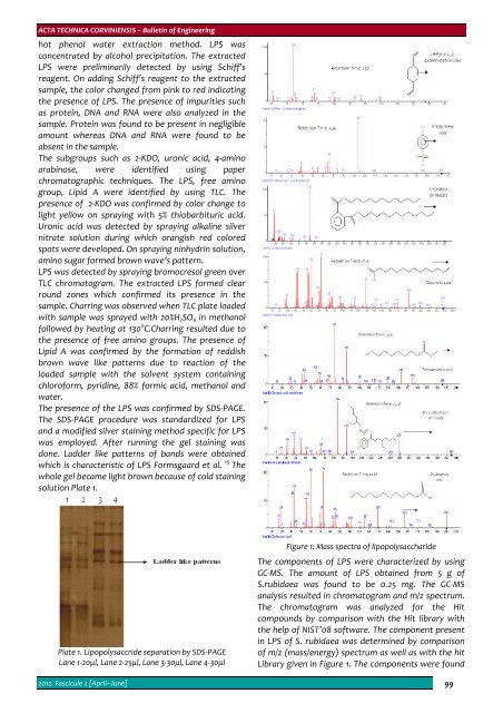
Figure 1: Mass spectra of lipopolysaccharide<br />
The components of LPS were characterized by using<br />
GC‐MS. The amount of LPS obtained from 5 g of<br />
S.rubidaea was found to be 0.25 mg. The GC‐MS<br />
analysis resulted in chromatogram and m/z spectrum.<br />
The chromatogram was analyzed for the Hit<br />
compounds by comparison with the Hit library with<br />
the help of NIST’08 software. The component present<br />
in LPS of S. rubidaea was determined by comparison<br />
of m/z (mass/energy) spectrum as well as with the hit<br />
Library given in Figure 1. The components were found<br />
2012. Fascicule 2 [April–June] 99