IJUP08 - Universidade do Porto
IJUP08 - Universidade do Porto
IJUP08 - Universidade do Porto
- TAGS
- universidade
- porto
- ijup.up.pt

Create successful ePaper yourself
Turn your PDF publications into a flip-book with our unique Google optimized e-Paper software.
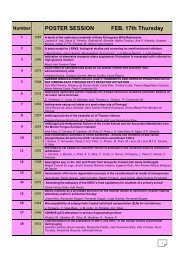
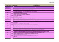
Anti-Angiogenic Effects of Ranibizumab and Bevacizumab in<br />
Age-Related Macular Degeneration: Effects on Human En<strong>do</strong>thelial<br />
Cells<br />
M. Ferreira-Pinto 1* , T. Taveira-Gomes 1* , A Carneiro 2 , M Falcão 2 , I. Azeve<strong>do</strong> 1 , F.<br />
Falcão-Reis 2 , R. Soares 1<br />
1 Department of Biochemistry, Faculty of Medicine of University of <strong>Porto</strong>, Portugal.<br />
2 Department od Ophthalmology, Faculty of Medicina of University of <strong>Porto</strong>, São João Hospital,<br />
Portugal<br />
* The two authors contributed equally to the project<br />
INTRODUCTION: Age-related macular degeneration (AMD) is the leading cause of<br />
irreversible blindness over 50 years of age in developed countries. This pathology is<br />
characterized by the development of abnormal choroidal blood vessels that proliferate<br />
through the Bruch's membrane, invading the subretinal space, beneath the macula, which<br />
causes severe and fast loss of vision. Angiogenesis, the formation of new blood vessels<br />
from pre-existing ones, is accomplished by a huge number of pro-angiogenic growth<br />
factors, such as vascular en<strong>do</strong>thelial growth factor (VEGF). Recent reports claiming that<br />
VEGF plays a crucial role in AMD-related angiogenesis rendered this growth factor a<br />
major therapeutic target. Ranibizumab and bevacizumab are two therapeutic agents already<br />
being used in the clinical pratice that block the action of VEGF. However, there are no<br />
studies comparing the relative efficacy and safety in AMD patients.<br />
OBJECTIVES: The purpose of the present study is to compare the effects of ranibizumab<br />
and bevacizumab in en<strong>do</strong>thelial cell growth, apoptosis, migration and in vitro capilary-like<br />
tubule formation.<br />
METHODS: Human Umbilical Vein En<strong>do</strong>thelial Cell (HUVEC) cultures were incubated<br />
with different concentrations of ranibizumab or bevacizumab within the <strong>do</strong>ses used in the<br />
clinic, or their excipients. HUVEC viability (by MTT), proliferation (by BrdU<br />
immunoassay), apoptosis (by TUNEL assay) and migration (using <strong>do</strong>uble-chamber assays)<br />
were performed. Evaluation of tubule-like structures formation was performed on matrigelcoated<br />
plaques incubated with ranibizumab, bevacizumab or their excipients.<br />
RESULTS: Neither bevacizumab nor ranibizumab presented cytotoxic effects, as evaluated<br />
by MTT assay. HUVEC proliferation was significantly reduced by the two agents, as<br />
compared to excipient-treated controls. Incubation with bevacizumab at five different<br />
concentrations led to increase apoptosis. Ranibizumab treatment resulted in increased<br />
apoptosis in the two higher concentrations tested. Cell migration was only affected by<br />
higher concentrations of both agents. Cell assembly into capillary-like structures was<br />
effectively <strong>do</strong>wnregulated by incubation with bevacizumab at the clinically used <strong>do</strong>se,<br />
whereas ranibizumab treatment resulted in decreased cord formation, although not<br />
reaching statistical significance.<br />
CONCLUSIONS: This study demonstrated that clinical <strong>do</strong>ses of bevacizumab and<br />
ranibizumab are able to prevent several steps of the angiogenic process in a distinct<br />
manner. Statistical analyses are being performed in order to elucidate the precise effects of<br />
each of the agents examined.<br />
157