

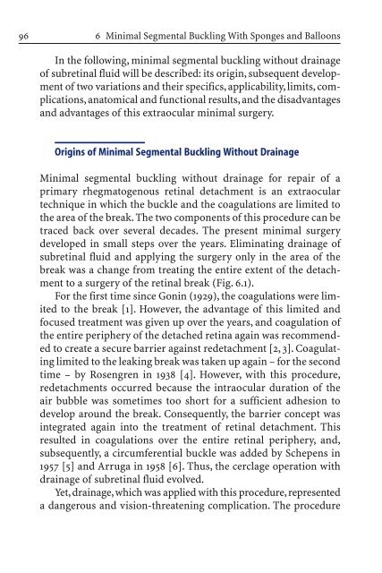
Primary Retinal Detachment
Primary Retinal Detachment
Primary Retinal Detachment

Create successful ePaper yourself
Turn your PDF publications into a flip-book with our unique Google optimized e-Paper software.
96<br />
6 Minimal Segmental Buckling With Sponges and Balloons<br />
In the following, minimal segmental buckling without drainage<br />
of subretinal fluid will be described: its origin, subsequent development<br />
of two variations and their specifics, applicability, limits, complications,<br />
anatomical and functional results, and the disadvantages<br />
and advantages of this extraocular minimal surgery.<br />
Origins of Minimal Segmental Buckling Without Drainage<br />
Minimal segmental buckling without drainage for repair of a<br />
primary rhegmatogenous retinal detachment is an extraocular<br />
technique in which the buckle and the coagulations are limited to<br />
the area of the break. The two components of this procedure can be<br />
traced back over several decades. The present minimal surgery<br />
developed in small steps over the years. Eliminating drainage of<br />
subretinal fluid and applying the surgery only in the area of the<br />
break was a change from treating the entire extent of the detachment<br />
to a surgery of the retinal break (Fig. 6.1).<br />
For the first time since Gonin (1929), the coagulations were limited<br />
to the break [1]. However, the advantage of this limited and<br />
focused treatment was given up over the years, and coagulation of<br />
the entire periphery of the detached retina again was recommended<br />
to create a secure barrier against redetachment [2, 3]. Coagulating<br />
limited to the leaking break was taken up again – for the second<br />
time – by Rosengren in 1938 [4]. However, with this procedure,<br />
redetachments occurred because the intraocular duration of the<br />
air bubble was sometimes too short for a sufficient adhesion to<br />
develop around the break. Consequently, the barrier concept was<br />
integrated again into the treatment of retinal detachment. This<br />
resulted in coagulations over the entire retinal periphery, and,<br />
subsequently, a circumferential buckle was added by Schepens in<br />
1957 [5] and Arruga in 1958 [6]. Thus, the cerclage operation with<br />
drainage of subretinal fluid evolved.<br />
Yet, drainage, which was applied with this procedure, represented<br />
a dangerous and vision-threatening complication. The procedure


