

Primary Retinal Detachment
Primary Retinal Detachment
Primary Retinal Detachment

Create successful ePaper yourself
Turn your PDF publications into a flip-book with our unique Google optimized e-Paper software.
182<br />
9 Repair of <strong>Primary</strong> <strong>Retinal</strong> <strong>Detachment</strong><br />
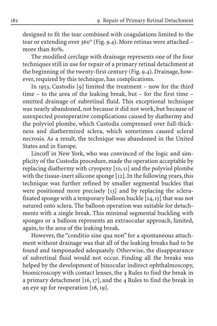
designed to fit the tear combined with coagulations limited to the<br />
tear or extending over 360° (Fig. 9.4). More retinas were attached –<br />
more than 80%.<br />
The modified cerclage with drainage represents one of the four<br />
techniques still in use for repair of a primary retinal detachment at<br />
the beginning of the twenty-first century (Fig. 9.4). Drainage, however,<br />
required by this technique, has complications.<br />
In 1953, Custodis [9] limited the treatment – now for the third<br />
time – to the area of the leaking break, but – for the first time –<br />
omitted drainage of subretinal fluid. This exceptional technique<br />
was nearly abandoned, not because it did not work, but because of<br />
unexpected postoperative complications caused by diathermy and<br />
the polyviol plombe, which Custodis compressed over full-thickness<br />
and diathermized sclera, which sometimes caused scleral<br />
necrosis. As a result, the technique was abandoned in the United<br />
States and in Europe.<br />
Lincoff in New York, who was convinced of the logic and simplicity<br />
of the Custodis procedure, made the operation acceptable by<br />
replacing diathermy with cryopexy [10, 11] and the polyviol plombe<br />
with the tissue-inert silicone sponge [12]. In the following years, this<br />
technique was further refined by smaller segmental buckles that<br />
were positioned more precisely [13] and by replacing the sclerafixated<br />
sponge with a temporary balloon buckle [14, 15] that was not<br />
sutured onto sclera. The balloon operation was suitable for detachments<br />
with a single break. This minimal segmental buckling with<br />
sponges or a balloon represents an extraocular approach, limited,<br />
again, to the area of the leaking break.<br />
However, the “conditio sine qua non” for a spontaneous attachment<br />
without drainage was that all of the leaking breaks had to be<br />
found and tamponaded adequately. Otherwise, the disappearance<br />
of subretinal fluid would not occur. Finding all the breaks was<br />
helped by the development of binocular indirect ophthalmoscopy,<br />
biomicroscopy with contact lenses, the 4 Rules to find the break in<br />
a primary detachment [16, 17], and the 4 Rules to find the break in<br />
an eye up for reoperation [18, 19].


