Mechanisms of Olfaction in Insects - ResearchSpace@Auckland ...
Mechanisms of Olfaction in Insects - ResearchSpace@Auckland ...
Mechanisms of Olfaction in Insects - ResearchSpace@Auckland ...

Create successful ePaper yourself
Turn your PDF publications into a flip-book with our unique Google optimized e-Paper software.
Functional characterisation <strong>of</strong> Epiphyas postvittana odorant receptor 1 44<br />
2.2.7 Data analysis<br />
The data collected from the calcium imag<strong>in</strong>g was analysed us<strong>in</strong>g MetaFluor Analyst<br />
v5.1 (Universal Imag<strong>in</strong>g Corporation). Circles were drawn with the region <strong>of</strong> <strong>in</strong>terest<br />
(ROI) tool around the perimeter <strong>of</strong> the cells <strong>in</strong> the field <strong>of</strong> view and also three<br />
background regions free <strong>of</strong> cells. The fluorescence data for the selected ROIs over the<br />
image acquisition period was obta<strong>in</strong>ed with the „calculate plot‟ function <strong>of</strong> the<br />
s<strong>of</strong>tware. The data from the three background ROIs was averaged and the ROI data<br />
from the cells was subtracted from this background, to m<strong>in</strong>imise differences <strong>in</strong> cell<br />
image backgrounds.<br />
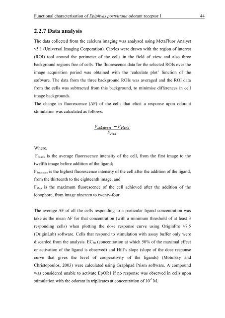
The change <strong>in</strong> fluorescence (∆F) <strong>of</strong> the cells that elicit a response upon odorant<br />
stimulation was calculated as follows:<br />
Where,<br />
FBlank is the average fluorescence <strong>in</strong>tensity <strong>of</strong> the cell, from the first image to the<br />
twelfth image before addition <strong>of</strong> the ligand;<br />
FSubstrate is the highest fluorescence <strong>in</strong>tensity <strong>of</strong> the cell after the addition <strong>of</strong> the ligand,<br />
from the thirteenth to the eighteenth image, and<br />
FMax is the maximum fluorescence <strong>of</strong> the cell achieved after the addition <strong>of</strong> the<br />
ionophore, from image n<strong>in</strong>eteen to twenty-four.<br />
The average ∆F <strong>of</strong> all the cells respond<strong>in</strong>g to a particular ligand concentration was<br />
take as the mean ∆F for that concentration (with a m<strong>in</strong>imum threshold <strong>of</strong> at least 3<br />
respond<strong>in</strong>g cells) when plott<strong>in</strong>g the dose response curve us<strong>in</strong>g Orig<strong>in</strong>Pro v7.5<br />
(Orig<strong>in</strong>Lab) s<strong>of</strong>tware. Cells that respond to stimulation with assay buffer only were<br />
discarded from the analysis. EC50 (concentration at which 50% <strong>of</strong> the maximal effect<br />
or activation <strong>of</strong> the ligand is observed) and Hill‟s slope (slope <strong>of</strong> the dose response<br />
curve that gives the level <strong>of</strong> cooperativity <strong>of</strong> the ligands) (Motulsky and<br />
Christopoulos, 2003) were calculated us<strong>in</strong>g Graphpad Prism s<strong>of</strong>tware. A compound<br />
was considered unable to activate EpOR1 if no response was observed <strong>in</strong> cells upon<br />
stimulation with the odorant <strong>in</strong> triplicates at concentration <strong>of</strong> 10 -5 M.