Mechanisms of Olfaction in Insects - ResearchSpace@Auckland ...
Mechanisms of Olfaction in Insects - ResearchSpace@Auckland ...
Mechanisms of Olfaction in Insects - ResearchSpace@Auckland ...

Create successful ePaper yourself
Turn your PDF publications into a flip-book with our unique Google optimized e-Paper software.
Functional characterisation <strong>of</strong> Epiphyas postvittana odorant receptor 1 46<br />
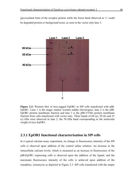
glycosylated form <strong>of</strong> the receptor prote<strong>in</strong> while the lower band observed at „c‟ could<br />
be degraded prote<strong>in</strong> or background noise, as seen <strong>in</strong> the vector only lane 3.<br />
Figure 2.2: Western blot <strong>of</strong> myc-tagged EpOR1 <strong>in</strong> Sf9 cells transfected with pIB-<br />
EpOR1. Lane 1 is the magic marker western ladder (Invitrogen), lane 2 is the pIB-<br />
EpOR1 prote<strong>in</strong> membrane fraction and lane 3 is the pIB-V5/His prote<strong>in</strong> membrane<br />
fraction from cells transfected with vector only. Three bands <strong>of</strong> 60 (a), 50 (b) and 25<br />
(c) kDa were observed <strong>in</strong> lane 2, the 50 kDa band correspond<strong>in</strong>g to the molecular<br />
weight <strong>of</strong> myc-EpOR1.<br />
2.3.1 EpOR1 functional characterisation <strong>in</strong> Sf9 cells<br />
In a typical calcium assay experiment, no change <strong>in</strong> fluorescence <strong>in</strong>tensity <strong>of</strong> the Sf9<br />
cells is observed upon addition <strong>of</strong> the control sal<strong>in</strong>e solution. An <strong>in</strong>crease <strong>in</strong> the<br />
<strong>in</strong>tracellular calcium levels, which is measured as an <strong>in</strong>crease <strong>in</strong> fluorescence <strong>of</strong> the<br />
pIB-EpOR1 express<strong>in</strong>g cells is observed upon the addition <strong>of</strong> the ligand, and the<br />
maximum fluorescence <strong>in</strong>tensity <strong>of</strong> the cells is achieved upon addition <strong>of</strong> the<br />
ionophore, ionomyc<strong>in</strong> as depicted <strong>in</strong> Figure 2.3. Sf9 cells transfected with the empty