

Eble JN, Sauter G., Epstein JI, Sesterhenn IA - iarc
Eble JN, Sauter G., Epstein JI, Sesterhenn IA - iarc
Eble JN, Sauter G., Epstein JI, Sesterhenn IA - iarc

Create successful ePaper yourself
Turn your PDF publications into a flip-book with our unique Google optimized e-Paper software.
A<br />
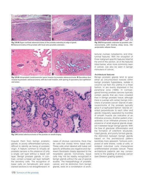
Fig. 3.12 A Organ-confined adenocarcinoma of the prostate extending to edge of gland.<br />
B Adenocarcinoma of the prostate with focal extra-prostatic extension.<br />
B<br />
Fig. 3.15 Extraprostatic extension by prostatic adenocarcinoma,<br />
with tracking along nerve, into<br />
periprostatic adipose tissue.<br />
cases of obvious carcinoma, there may<br />
be cells that closely mimic basal cells.<br />
These cells when labeled with basal cell<br />
specific antibodies are negative and represent<br />
fibroblasts closely apposed to the<br />
neoplastic glands. Conversely, basal<br />
cells may not be readily recognized in<br />
benign glands without the use of special<br />
studies. The histopathology of prostatic<br />
cancer, and its distinction from benign<br />
glands, rests on a constellation of architectural,<br />
nuclear, cytoplasmic, and intraluminal<br />
features. With the exception of<br />
three malignant specific features listed at<br />
the end of this section, all of the features<br />
listed below, while more commonly seen<br />
in cancer, can also be seen in benign<br />
mimickers of cancer.<br />
A<br />
Fig. 3.13 A Intraprostatic lymphovascular space invasion by prostatic adenocarcinoma. B Ejaculatory duct<br />
invasion by prostatic adenocarcinoma, with duct wall invasion, with sparing of ejaculatory duct epithelium<br />
and lumen.<br />
Fig. 3.14 Limited adenocarcinoma of the prostate on needle biopsy.<br />
tinguish them from benign prostatic<br />
glands, to poorly differentiated tumours,<br />
difficult to identify as being of prostatic<br />
origin. A feature common to virtually all<br />
prostate cancers is the presence of only<br />
a single cell type without a basal cell<br />
layer. Benign prostate glands, in contrast,<br />
contain a basal cell layer beneath<br />
the secretory cells. The recognition of<br />
basal cells on hematoxylin and eosin<br />
stained sections is not straightforward. In<br />
B<br />
Architectural features<br />
Benign prostatic glands tend to grow<br />
either as circumscribed nodules within<br />
benign prostatic hyperplasia, radiate in<br />
columns out from the urethra in a linear<br />
fashion, or are evenly dispersed in the<br />
peripheral zone {1685}. In contrast,<br />
gland-forming prostate cancers typically<br />
contain glands that are more crowded<br />
than in benign prostatic tissue, although<br />
there is overlap with certain benign mimickers<br />
of prostate cancer. Glands of adenocarcinoma<br />
of the prostate typically<br />
grow in a haphazard fashion. Glands oriented<br />
perpendicular to each other and<br />
glands irregularly separated by bundles<br />
of smooth muscle are indicative of an<br />
infiltrative process. Another pattern characteristic<br />
of an infiltrative process is the<br />
presence of small atypical glands situated<br />
in between larger benign glands. With<br />
the loss of glandular differentiation and<br />
the formation of cribriform structures,<br />
fused glands, and poorly formed glands,<br />
the distinction between benign glands<br />
based on the architectural pattern<br />
becomes more apparent. Tumours composed<br />
of solid sheets, cords of cells, or<br />
isolated individual cells characterize<br />
undifferentiated prostate cancer. These<br />
architectural patterns are key components<br />
to the grading of prostate cancer<br />
(see Gleason grading system).<br />
170 Tumours of the prostate


