

Eble JN, Sauter G., Epstein JI, Sesterhenn IA - iarc
Eble JN, Sauter G., Epstein JI, Sesterhenn IA - iarc
Eble JN, Sauter G., Epstein JI, Sesterhenn IA - iarc

Create successful ePaper yourself
Turn your PDF publications into a flip-book with our unique Google optimized e-Paper software.
Metanephric adenoma and metanephric<br />
adenofibroma<br />
J.N. <strong>Eble</strong><br />
D.J. Grignon<br />
H. Moch<br />
Definition<br />
Metanephric adenoma is a highly cellular<br />
epithelial tumour composed of small, uniform,<br />
embryonic-appearing cells.<br />
ICD-O codes<br />
Metanephric adenoma 8325/0<br />
Metanephric adenofibroma 9013/0<br />
Metanephric adenosarcoma 8933/3<br />
Epidemiology<br />
Metanephric adenoma occurs in children<br />
and adults, most commonly in the fifth<br />
and sixth decades. There is a 2:1 female<br />
preponderance {561}. Patients with<br />
metanephric adenofibroma have ranged<br />
from 5 months to 36 years (median = 30<br />
months) {120}. There is a 2:1 ratio of males<br />
to females. A single case of high grade sarcoma<br />
arising in association with<br />
metanephric adenoma (metanephric<br />
adenosarcoma) has been reported {2072}.<br />
Clinical features<br />
Approximately 50% of metanephric adenoma<br />
are incidental findings with others<br />
presenting with polycythemia, abdominal<br />
or flank pain, mass, or hematuria.<br />
Presenting symptoms of metanephric<br />
adenofibroma have included polycythemia<br />
or hematuria; some have been<br />
incidental findings. Arroyo et al. {120}<br />
described several cases in which either<br />
Wilms tumour or carcinoma occurred in<br />
association with metanephric adenofibroma.<br />
Other than one patient with<br />
regional metastases from the carcinoma,<br />
these patients have had no progression.<br />
Macroscopy<br />
Metanephric adenomas range widely in<br />
size; most have been 30 to 60 mm in<br />
diameter {561}. Multifocality is uncommon.<br />
The tumours are typically well circumscribed<br />
but not encapsulated. The<br />
cut surfaces vary from grey to tan to yellow<br />
and may be soft or firm.<br />
Foci of haemorrhage and necrosis are<br />
common; calcification is present in<br />
approximately 20%,and small cysts in<br />
10% {561,1237}.<br />
Metanephric adenofibromas are typically<br />
solitary tan partially cystic masses with<br />
indistinct borders {120}.<br />
Histopathology<br />
Metanephric adenoma is a highly cellular<br />
tumour composed of tightly packed<br />
small, uniform, round acini with an<br />
embryonal appearance. Since the acini<br />
and their lumens are small, at low magnification<br />
this pattern may be mistaken for<br />
a solid sheet of cells. Long branching<br />
and angulated tubular structures also are<br />
common. The stroma ranges from inconspicuous<br />
to a loose oedematous stroma.<br />
Fig. 1.57 Metanephric adenoma.<br />
Hyalinized scar and focal osseous metaplasia<br />
of the stroma are present in 10-<br />
20% of tumours {561}. Approximately<br />
50% of tumours contain papillary structures,<br />
usually consisting of tiny cysts into<br />
which protrude blunt papillae reminiscent<br />
of immature glomeruli. Psammoma<br />
bodies are common and sometimes<br />
numerous. The junction with the kidney is<br />
usually sharp and without a pseudocapsule.<br />
The cells of metanephric adenoma<br />
are monotonous, with small, uniform<br />
nuclei and absent or inconspicuous<br />
nucleoli. The nuclei are only a little larger<br />
than those of lymphocytes and are round<br />
or oval with delicate chromatin. The cytoplasm<br />
is scant and pale or light pink.<br />
Mitotic figures are absent or rare.<br />
Metanephric adenofibroma is a compos-<br />
A<br />
B<br />
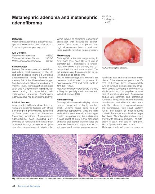
Fig. 1.58 Metanephric adenoma. A Well circumscribed tumour without encapsulation. B Complicated ductal architecture with psammoma bodies.<br />
44 Tumours of the kidney


