- Page 1 and 2: World Health Organization Classific
- Page 3 and 4: This volume was produced in collabo
- Page 5 and 6: Contents 1 Tumours of the kidney 9
- Page 7 and 8: CHAPTER 1 Tumours of the Kidney Can
- Page 9 and 10: TNM classification of renal cell ca
- Page 11 and 12: one quarter of kidney cancers in bo
- Page 13 and 14: Familial renal cell carcinoma M.J.
- Page 15 and 16: Table 1.02 Genotype - phenotype cor
- Page 17 and 18: A B Fig. 1.11 A Multiple cutaneous
- Page 19 and 20: A B Fig. 1.14 Birt-Hogg-Dubé syndr
- Page 21 and 22: Clear cell renal cell carcinoma D.J
- Page 23 and 24: Fig. 1.20 Clear cell renal cell car
- Page 25 and 26: Papillary renal cell carcinoma B. D
- Page 27 and 28: A B Fig. 1.29 Papillary renal cell
- Page 29 and 30: Fig. 1.33 Chromophobe RCC with sarc
- Page 31 and 32: Carcinoma of the collecting ducts o
- Page 33 and 34: Renal medullary carcinoma C.J. Davi
- Page 35 and 36: Renal carcinomas associated with Xp
- Page 37 and 38: Renal cell carcinoma associated wit
- Page 39 and 40: Papillary adenoma of the kidney J.N
- Page 41 and 42: of this tumour. Microscopic extensi
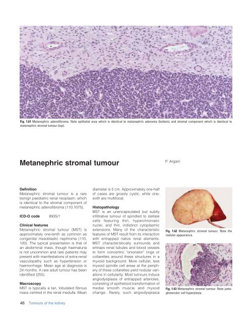
- Page 43: A B Fig. 1.59 Metanephric adenoma.
- Page 47 and 48: peritumoural fibrous pseudocapsule.
- Page 49 and 50: increases in prevalence to approxim
- Page 51 and 52: Nephrogenic rests and nephroblastom
- Page 53 and 54: Cystic partially differentiated nep
- Page 55 and 56: A B Fig. 1.80 Clear cell sarcoma of
- Page 57 and 58: A B Fig. 1.85 Rhabdoid tumour of th
- Page 59 and 60: transcription factor is fused to th
- Page 61 and 62: Leiomyosarcoma S.M. Bonsib Definiti
- Page 63 and 64: Angiomyolipoma G. Martignoni M.B. A
- Page 65 and 66: melanocytic and smooth muscle marke
- Page 67 and 68: Multinucleated and enlarged ganglio
- Page 69 and 70: Haemangioma P. Tamboli Definition H
- Page 71 and 72: Fig. 1.107 Juxtaglomerular cell tum
- Page 73 and 74: Intrarenal schwannoma I. Alvarado-C
- Page 75 and 76: Mixed epithelial and stromal tumour
- Page 77 and 78: Synovial sarcoma of the kidney J.Y.
- Page 79 and 80: Renal carcinoid tumour L.R. Bégin
- Page 81 and 82: Primitive neuroectodermal tumour (E
- Page 83 and 84: Paraganglioma / Phaeochromocytoma P
- Page 85 and 86: Leukaemia A. Orazi Interstitial inf
- Page 87 and 88: WHO histological classification of
- Page 89 and 90: TNM classification of carcinomas of
- Page 91 and 92: The risk of bladder cancer goes dow
- Page 93 and 94: Tumour spread and staging Urinary b
- Page 95 and 96:
feature in such patients undergoing
- Page 97 and 98:
A Fig. 2.12 Infiltrative urothelial
- Page 99 and 100:
A Fig. 2.15 A Infiltrating urotheli
- Page 101 and 102:
A B Fig. 2.19 A Infiltrative urothe
- Page 103 and 104:
Fig. 2.23 Infiltrative urothelial c
- Page 105 and 106:
ing systems have been proposed on t
- Page 107 and 108:
Non-invasive urothelial tumours G.
- Page 109 and 110:
chronically inflamed urothelium and
- Page 111 and 112:
Inverted papilloma G. Sauter Defini
- Page 113 and 114:
muscle invasive disease, but there
- Page 115 and 116:
Fig. 2.42 Non-invasive urothelial n
- Page 117 and 118:
A Fig. 2.45 Non-invasive urothelial
- Page 119 and 120:
for DBCCR1 silencing {984,2476}. Th
- Page 121 and 122:
Squamous cell carcinoma D.J. Grigno
- Page 123 and 124:
Fig. 2.51 Squamous cell carcinoma.
- Page 125 and 126:
Adenocarcinoma A.G. Ayala P. Tambol
- Page 127 and 128:
A B Fig. 2.61 A Adenocarcinoma in s
- Page 129 and 130:
A B Fig. 2.65 Intramural urachal ca
- Page 131 and 132:
Müllerian origin is postulated for
- Page 133 and 134:
A Fig. 2.69 Small cell carcinoma. A
- Page 135 and 136:
Carcinoid L. Cheng Definition Carci
- Page 137 and 138:
Leiomyosarcoma J. Cheville Definiti
- Page 139 and 140:
Osteosarcoma L. Guillou Definition
- Page 141 and 142:
Leiomyoma J. Cheville Definition A
- Page 143 and 144:
Haemangioma L. Cheng Definition Hae
- Page 145 and 146:
Metastatic tumours and secondary ex
- Page 147 and 148:
Tumours of the renal pelvis and ure
- Page 149 and 150:
nodular, ulcerative or infiltrative
- Page 151 and 152:
Tumours of the urethra F. Hofstädt
- Page 153 and 154:
usually show enteric, colloid or si
- Page 155 and 156:
CHAPTER 3X Tumours of of the the Pr
- Page 157 and 158:
TNM classification of carcinomas of
- Page 159 and 160:
contrast, mortality among migrants
- Page 161 and 162:
Fig. 3.06 Transrectal ultrasound of
- Page 163 and 164:
PSA-related diagnostic strategies.
- Page 165 and 166:
A B C Fig. 3.10 A,B Section of pros
- Page 167 and 168:
pale-clear, similar to benign gland
- Page 169 and 170:
A B Fig. 3.20 A, B Adenocarcinoma w
- Page 171 and 172:
prostate adenocarcinomas exhibit AR
- Page 173 and 174:
A Fig. 3.27 A, B Foamy gland adenoc
- Page 175 and 176:
A B Fig. 3.32 A Sarcomatoid carcino
- Page 177 and 178:
A B Fig. 3.37 A Gleason score 1+1=2
- Page 179 and 180:
A B Fig. 3.43 A Prostate cancer Gle
- Page 181 and 182:
Fig. 3.48 Heat map-nature. From S.M
- Page 183 and 184:
Fig. 3.51 Prostate cancer. Major su
- Page 185 and 186:
many stage T1b cancers. Stage T2 Mo
- Page 187 and 188:
Fig. 3.58 Patterns of seminal vesic
- Page 189 and 190:
Prostatic intraepithelial neoplasia
- Page 191 and 192:
A B Fig. 3.63 A Micropapillary high
- Page 193 and 194:
A B Fig. 3.68 A Ductal carcinoma in
- Page 195 and 196:
Ductal adenocarcinoma X.J. Yang L.
- Page 197 and 198:
Papillary pattern can be seen in bo
- Page 199 and 200:
A B Fig. 3.74 A Inflammation withou
- Page 201 and 202:
Squamous neoplasms T.H. Van der Kwa
- Page 203 and 204:
Neuroendocrine tumours P.A. di Sant
- Page 205 and 206:
Mesenchymal tumours J. Cheville F.
- Page 207 and 208:
Fig. 3.89 Sarcoma of the prostate.
- Page 209 and 210:
Miscellaneous tumours P.H. Tan L. C
- Page 211 and 212:
A Fig. 3.96 A Adenocarcinoma of the
- Page 213 and 214:
WHO histological classification of
- Page 215 and 216:
Introduction F.K. Mostofi I.A. Sest
- Page 217 and 218:
wartime birth cohorts illustrate th
- Page 219 and 220:
Similar tumours as those of group 1
- Page 221 and 222:
crucial for the development of this
- Page 223 and 224:
A B Fig. 4.09 Spermatocytic seminom
- Page 225 and 226:
A B Fig. 4.14 Intratubular germ cel
- Page 227 and 228:
A B Fig. 4.19 Seminoma. A Typical s
- Page 229 and 230:
Fig. 4.24 Seminoma. Vascular invasi
- Page 231 and 232:
Fig. 4.28 Spermatocytic seminoma wi
- Page 233 and 234:
A B Fig. 4.33 Embryonal carcinoma.
- Page 235 and 236:
A B Fig. 4.38 Yolk sac tumour. A En
- Page 237 and 238:
have cytoplasmic lacunae that conta
- Page 239 and 240:
A Fig. 4.46 Teratoma. A Longitudina
- Page 241 and 242:
A B Fig. 4.51 A Cut surface of derm
- Page 243 and 244:
Fig. 4.54 Mixed germ cell tumour. L
- Page 245 and 246:
Sex cord / gonadal stromal tumours
- Page 247 and 248:
A B C Fig. 4.67 Malignant Leydig ce
- Page 249 and 250:
A Fig. 4.73 A Sertoli cell tumour.
- Page 251 and 252:
ICD-O codes Granulosa cell tumour 8
- Page 253 and 254:
ICD-O code 8592/1 Clinical features
- Page 255 and 256:
A B Fig. 4.83 Germ cell-sex cord/go
- Page 257 and 258:
A Fig. 4.85 A, B Brenner tumour of
- Page 259 and 260:
typical grade III follicular morpho
- Page 261 and 262:
testicular parenchyma and tumour te
- Page 263 and 264:
A B the surgical scar and adjacent
- Page 265 and 266:
Immunoprofile Ordóñez and associa
- Page 267 and 268:
often have a grey-white cut surface
- Page 269 and 270:
Fig. 4.111 Angiomyofibroblastoma-li
- Page 271 and 272:
A B Fig. 4.119 Embryonal rhabdomyos
- Page 273 and 274:
Table 4.05 Secondary tumours of the
- Page 275 and 276:
WHO histological classification of
- Page 277 and 278:
A Fig. 5.04 A, B Squamous cell carc
- Page 279 and 280:
deformed by an exophytic mass. In s
- Page 281 and 282:
A B C Fig. 5.12 A Warty (condylomat
- Page 283 and 284:
A Fig. 5.20 Bowenoid papulosis. A,
- Page 285 and 286:
three had been circumcized by 9 yea
- Page 287 and 288:
Mesenchymal tumours J.F. Fetsch M.
- Page 289 and 290:
Fig. 5.31 Neurofibroma of the penis
- Page 291 and 292:
oectodermal tumour/Ewing sarcoma [p
- Page 293 and 294:
Secondary tumours of the penis C.J.
- Page 295 and 296:
Dr John N. EBLE* Dept. of Pathology
- Page 297 and 298:
Dr Ricardo PANIAGUA Department of C
- Page 299 and 300:
Source of charts and photographs 1.
- Page 301 and 302:
References 1. Anon. (1955). Case re
- Page 303 and 304:
115. Ariel I, Sughayer M, Fellig Y,
- Page 305 and 306:
246. Birt AR, Hogg GR, Dube WJ (197
- Page 307 and 308:
374. Cardone G, Malventi M, Roffi M
- Page 309 and 310:
502. Corti B, Carella R, Gabusi E,
- Page 311 and 312:
633. Donhuijsen K, Schmidt U, Richt
- Page 313 and 314:
768. Fischer J, Palmedo G, von Knob
- Page 315 and 316:
900. Goedert JJ, Cote TR, Virgo P,
- Page 317 and 318:
1028. Hartmann A, Dietmaier W, Hofs
- Page 319 and 320:
1162. Iezzoni JC, Fechner RE, Wong
- Page 321 and 322:
1293. Keetch DW, Catalona WJ (1995)
- Page 323 and 324:
1424. Ladanyi M, Lui MY, Antonescu
- Page 325 and 326:
1548. Lopez-Beltran A, Croghan GA,
- Page 327 and 328:
1681. McLaughlin JK, Silverman DT,
- Page 329 and 330:
1816. Moudouni SM, En-Nia I, Rioux-
- Page 331 and 332:
1945. Ohori M, Wheeler TM, Kattan M
- Page 333 and 334:
2070. Phillips G, Kumari-Subaiya S,
- Page 335 and 336:
2199. Riou G, Barrois M, Prost S, T
- Page 337 and 338:
2327. Schmidt L, Junker K, Weirich
- Page 339 and 340:
2450. Smith G, Elton RA, Beynon LL,
- Page 341 and 342:
2572. Tamboli P, Mohsin SK, Hailema
- Page 343 and 344:
2693. van Echten J, Timmer A, van d
- Page 345 and 346:
2816. Wick MR, Berg LC, Hertz MI (1
- Page 347 and 348:
2937. Zhuang Z, Park WS, Pack S, Sc
- Page 349 and 350:
CCNB, 226 CCND1, 105, 108 CCND2, 22
- Page 351 and 352:
J Juvenile type granulosa cell tumo
- Page 353 and 354:
Promoter methylation, 21 Prostate s