in vitro PHARMACOLOGY 2011 CATALOG - Cerep
in vitro PHARMACOLOGY 2011 CATALOG - Cerep
in vitro PHARMACOLOGY 2011 CATALOG - Cerep

Create successful ePaper yourself
Turn your PDF publications into a flip-book with our unique Google optimized e-Paper software.
123<br />
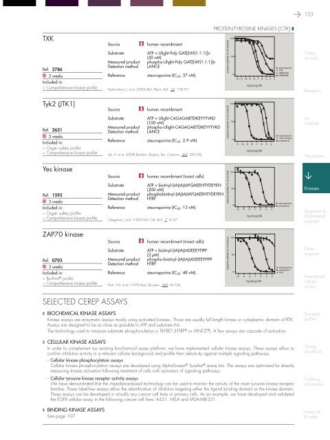
TXK<br />
Ref. 2786<br />
Q 3 weeks<br />
Included <strong>in</strong>:<br />
Comprehensive k<strong>in</strong>ase profile<br />
Source<br />
human recomb<strong>in</strong>ant<br />
Substrate<br />
ATP + Ulight-Poly GAT[EAY(1:1:1)]n<br />
(50 nM)<br />
Measured product phospho-Ulight-Poly GAT[EAY(1:1:1)]n<br />
Detection method LANCE<br />
Reference<br />
staurospor<strong>in</strong>e (IC 50 : 57 nM)<br />
Kashiwakura, J. et al. (2002) Biol. Pharm. Bull., 25: 718-721.<br />
prote<strong>in</strong>-tyros<strong>in</strong>e k<strong>in</strong>ases [Ctk] ❚<br />
<br />
<br />
<br />
<br />
<br />
<br />
<br />
<br />
<br />
<br />
<strong>Cerep</strong><br />
services<br />
Receptors<br />
Tyk2 (JTK1)<br />
Ref. 2621<br />
Q 3 weeks<br />
Included <strong>in</strong>:<br />
Organ safety profile<br />
Comprehensive k<strong>in</strong>ase profile<br />
Source<br />
human recomb<strong>in</strong>ant<br />
Substrate<br />
ATP + Ulight-CAGAGAIETDKEYYTVKD<br />
(100 nM)<br />
Measured product phospho-Ulight-CAGAGAIETDKEYYTVKD<br />
Detection method LANCE<br />
Reference<br />
staurospor<strong>in</strong>e (IC 50 : 2.9 nM)<br />
Ide, H. et al. (2008) Biochem. Biophys. Res. Commun., 369: 292-296.<br />
<br />
<br />
<br />
<br />
<br />
<br />
<br />
<br />
<br />
<br />
<br />
<br />
<br />
<br />
<br />
<br />
<br />
<br />
<br />
Ion<br />
channels<br />
Transporters<br />
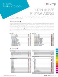
Yes k<strong>in</strong>ase<br />
Ref. 1595<br />
Q 3 weeks<br />
Included <strong>in</strong>:<br />
Organ safety profile<br />
Comprehensive k<strong>in</strong>ase profile<br />
Source<br />
human recomb<strong>in</strong>ant (<strong>in</strong>sect cells)<br />
Substrate<br />
ATP + biot<strong>in</strong>yl-βAβAβAYQAEENTYDEYEN<br />
(200 nM)<br />
Measured product phosphobiot<strong>in</strong>yl-βAβAβAYQAEENTYDEYEN<br />
Detection method HTRF<br />
Reference<br />
staurospor<strong>in</strong>e (IC 50 : 13 nM)<br />
Sukegawa J. et al. (1987) Mol. Cell. Biol., 7: 41-47.<br />
enzyme activity (% of control)<br />
<br />
<br />
100<br />
<br />
<br />
50<br />
<br />
<br />
staurospor<strong>in</strong>e<br />
0<br />
piceatannol<br />
-10 -9 -8 -7 -6 -5 -4<br />
<br />
<br />
log [drug] (M)<br />
<br />
K<strong>in</strong>ases<br />
Epigenetic &<br />
DNA-related<br />
enzymes<br />
ZAP70 k<strong>in</strong>ase<br />
Ref. 0705<br />
Q 3 weeks<br />
Included <strong>in</strong>:<br />
BioPr<strong>in</strong>t ® profile<br />
Comprehensive k<strong>in</strong>ase profile<br />
Source<br />
human recomb<strong>in</strong>ant (<strong>in</strong>sect cells)<br />
Substrate<br />
ATP + biot<strong>in</strong>yl-βAβAβADEEEYFIPP<br />
(2 µM)<br />
Measured product phospho-biot<strong>in</strong>yl-βAβAβADEEEYFIPP<br />
Detection method HTRF<br />
Reference<br />
staurospor<strong>in</strong>e (IC 50 : 48 nM)<br />
Park, Y.M. et al. (1999) Anal. Biochem., 269: 94-104.<br />
<br />
<br />
<br />
<br />
<br />
<br />
<br />
<br />
<br />
Other<br />
enzymes<br />
Specialized<br />
cellular<br />
assays<br />
selected cerep assays<br />
❚ Biochemical k<strong>in</strong>ase assays<br />
K<strong>in</strong>ase assays are enzymatic assays mostly us<strong>in</strong>g activated k<strong>in</strong>ases. Those are usually full length k<strong>in</strong>ase or cytoplasmic doma<strong>in</strong> of RTK.<br />
Assays are designed to be as close as possible to ATP and substrate Km.<br />
The technology used to measure substrate phosphorylation is TR-FRET (HTRF ® or LANCE ® ). A few assays are cascade of activation.<br />
❚ Cellular k<strong>in</strong>ase assays<br />
In order to complement our exist<strong>in</strong>g biochemical assay platform, we have implemented cellular k<strong>in</strong>ase assays. These assays allow to<br />
confirm <strong>in</strong>hibitors activity <strong>in</strong> a relevant cellular background and profile their selectivity aga<strong>in</strong>st multiple signal<strong>in</strong>g pathways.<br />
- Cellular k<strong>in</strong>ase phosphorylation assays<br />
Cellular k<strong>in</strong>ase phosphorylation assays are developed us<strong>in</strong>g AlphaScreen ® Surefire ® assay kits. The assays are optimized for directly<br />
measur<strong>in</strong>g k<strong>in</strong>ase activation follow<strong>in</strong>g treatment of cells with activators of signal<strong>in</strong>g pathways.<br />
- Cellular tyros<strong>in</strong>e k<strong>in</strong>ase receptor activity assays<br />
We have demonstrated that the impedance-based technology can be used to monitor the activity of the ma<strong>in</strong> tyros<strong>in</strong>e k<strong>in</strong>ase receptor<br />
families. These label-free assays allow the identification of <strong>in</strong>hibitors target<strong>in</strong>g either the ligand b<strong>in</strong>d<strong>in</strong>g doma<strong>in</strong> or the k<strong>in</strong>ase doma<strong>in</strong>.<br />
These assays can be developed <strong>in</strong> virtually any cancer cell l<strong>in</strong>es or primary cells. As an example, we have developed and validated<br />
the EGFR cellular assay <strong>in</strong> the follow<strong>in</strong>g cancer cell l<strong>in</strong>es: A431, HELA and MDA-MB-231.<br />
❚ B<strong>in</strong>d<strong>in</strong>g k<strong>in</strong>ase assays<br />
See page 107.<br />
Standard<br />
profiles<br />
Test<strong>in</strong>g<br />
conditions<br />
Order<strong>in</strong>g<br />
<strong>in</strong>formation<br />
Assay list<br />
& <strong>in</strong>dex