in vitro PHARMACOLOGY 2011 CATALOG - Cerep
in vitro PHARMACOLOGY 2011 CATALOG - Cerep
in vitro PHARMACOLOGY 2011 CATALOG - Cerep

Create successful ePaper yourself
Turn your PDF publications into a flip-book with our unique Google optimized e-Paper software.
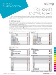
70 <strong>in</strong> <strong>vitro</strong> pharmacology <strong>2011</strong> catalog<br />
❚ seroton<strong>in</strong><br />
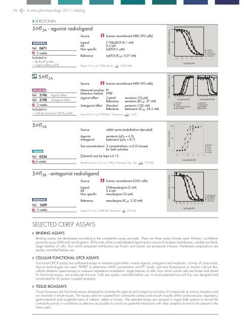
5-HT 2A - agonist radioligand<br />
b<strong>in</strong>d<strong>in</strong>g<br />
Ref. 0471<br />
Q 3 weeks<br />
Included <strong>in</strong>:<br />
BioPr<strong>in</strong>t ® profile<br />
Organ safety profile<br />
Source<br />
Ligand<br />
Kd<br />
Non specific<br />
Reference<br />
human recomb<strong>in</strong>ant (HEK-293 cells)<br />
[ 125 I](±)DOI (0.1 nM)<br />
0.3 nM<br />
(±)DOI (1 µM)<br />
(±)DOI (IC 50 : 0.27 nM)<br />
Bryant, H.U. et al. (1996) Life Sci., 15: 1259-1268.<br />
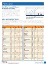
specific b<strong>in</strong>d<strong>in</strong>g (% of control)<br />
100<br />
50<br />
0<br />
-11 -10 -9 -8 -7 -6<br />
log [drug] (M)<br />
(+)DOI -<br />
seroton<strong>in</strong><br />
mesulerg<strong>in</strong>e<br />
ketanser<strong>in</strong><br />
5-HT 2A<br />
cellul ar<br />
Ref. 3196<br />
Ref. 3198<br />
Q 3 weeks<br />
Included <strong>in</strong>:<br />
Agonist effect<br />
Antagonist effect<br />
Cellular functional GPCR profile<br />
Source<br />
human recomb<strong>in</strong>ant (HEK-293 cells)<br />
Measured product IP 1<br />
Detection method HTRF<br />
Agonist effect Control seroton<strong>in</strong> (10 µM)<br />
Reference seroton<strong>in</strong> (EC 50 : 27 nM)<br />
Antagonist effect Stimulant seroton<strong>in</strong> (100 nM)<br />
Reference ketanser<strong>in</strong> (IC 50 : 24.2 nM)<br />
Porter, R.H.P. et al.(1999) Brit. J. Pharmacol., 128: 13-20.<br />
Ca 2+ mobilization (% of control)<br />
100<br />
50<br />
0<br />
-12 -11 -10 -9 -8 -7 -6 -5 -4<br />
log [agonist] (M)<br />
seroton<strong>in</strong><br />
(±)DOI<br />
5-methoxytryptam<strong>in</strong>e<br />
BW 723C86<br />
100<br />
50<br />
0<br />
-12 -11 -10 -9 -8 -7 -6 -5 -4<br />
log [antagonist] (M)<br />
ketanser<strong>in</strong><br />
mesulerg<strong>in</strong>e<br />
SB 206553<br />
SB 204741<br />
5-HT 2A<br />
tissue<br />
Ref. 0334<br />
Q 4 weeks<br />
Source<br />
rabbit aorta (endothelium-denuded)<br />
Agonist seroton<strong>in</strong> (pD 2 = 6.5)<br />
Antagonist ketanser<strong>in</strong> (pA 2 = 8.7)<br />
Test concentrations 3 concentrations, n=2 (2 tissues)<br />
for both activities<br />
[Solvent] must be kept ≤ 0.1%<br />
R<strong>in</strong>aldi-Carmona, M. et al. (1992) J. Pharmacol. Exp. Ther., 262: 759-768.<br />
tension (% of max.)<br />
-12 -11<br />
100<br />
50<br />
-10 -9 -8 -7 -6 -5<br />
-9 -8 -7 -6 -5<br />
-10 -4<br />
0<br />
-8 -7 -6 -5 -4 -3<br />
log [agonist] (M)<br />
ketanser<strong>in</strong><br />
none<br />
3 nM<br />
10 nM<br />
30 nM<br />
5-HT 2B - antagonist radioligand<br />
Source<br />
human recomb<strong>in</strong>ant (CHO cells)<br />
Ligand<br />
[ 3 H]mesulerg<strong>in</strong>e (2 nM)<br />
Kd<br />
2.4 nM<br />
Non specific mesulerg<strong>in</strong>e (10 µM)<br />
b<strong>in</strong>d<strong>in</strong>g<br />
Reference mesulerg<strong>in</strong>e (IC 50 : 2.35 nM)<br />
Ref. 1609<br />
Q 3 weeks<br />
Kursar, J.D. et al. (1994) Mol. Pharmacol., 46: 227-234.<br />
specific b<strong>in</strong>d<strong>in</strong>g (% of control)<br />
100<br />
50<br />
0<br />
-11<br />
-10 -9 -8 -7 -6 -5 -4<br />
log [drug] (M)<br />
mesulerg<strong>in</strong>e<br />
SB 206553<br />
SB 204741<br />
ketanser<strong>in</strong><br />
selected cerep assays<br />
❚ b<strong>in</strong>d<strong>in</strong>g assays<br />
-10 -9 -8 -7 -6 -5 -4 -3<br />
-11<br />
-10 -9 -8 -7 -6 -5 -4 -3<br />
-12 -11 -10 -9 -8 -7 -6 -5 -4<br />
B<strong>in</strong>d<strong>in</strong>g assays are developed accord<strong>in</strong>g to the competition assay pr<strong>in</strong>ciple. There are three assay formats -13 -12 used: -11 -10filtration, -9 -8 -7 -6sc<strong>in</strong>tillation<br />
-5<br />
-10 -9 -8 -7 -6<br />
proximity assay (SPA) and centrifugation. All formats utilize a radiolabeled ligand and a source of receptor (membranes, soluble/purified).<br />
Large batches of cells, from which prepared membranes are frozen and stored, are produced <strong>in</strong>-house. Membrane preparations are<br />
-10 -9 -8 -7 -6 -5 -4<br />
quality controlled before use.<br />
-12 -11 -10 -9 -8 -7 -6 -5 -4 -3<br />
❚ cellular Functional GPCR assays<br />
-12 -11 -10 -9 -8 -7 -6 -5<br />
Functional GPCR assays are cell-based assays to measure agonist-like, <strong>in</strong>verse agonist, antagonist and modulator<br />
-11 -10 -9<br />
activity<br />
-8 -7 -6of -5compounds.<br />
-4<br />
Various technologies are used: TR-FRET to determ<strong>in</strong>e cAMP concentration and IP1 levels, real time fluorescence to monitor calcium flux,<br />
cellular dielectric spectroscopy to measure impedance modulation. Large batches of cells, from which whole cells are frozen and stored<br />
for functional assays, are produced <strong>in</strong>-house. Cells are quality controlled before use. A novel patented host cell l<strong>in</strong>e was designed and<br />
constructed for Gi prote<strong>in</strong> coupled receptors.<br />
❚ tissue bioassays<br />
Tissue bioassays are functional assays designed to evaluate the agonist and antagonist activities of compounds at various receptors and<br />
ion channels <strong>in</strong> whole tissues. The tissues used are isolated from contractile cardiac and smooth muscles of the cardiovascular, respiratory,<br />
gastro<strong>in</strong>test<strong>in</strong>al and urogenital tracts of rodents, rabbit or human. The selected tissues are assayed <strong>in</strong> organ bath systems to record the<br />
contractile activity <strong>in</strong> conditions as selective as possible to avoid any potential <strong>in</strong>teraction with other receptors known to be present <strong>in</strong> the<br />
tissue used.